Domain 4: Surgical Correction Options for Failed Childhood Clubfoot Outcomes in Adults
Overview
Adults with painful, deformed feet resulting from failed or incomplete childhood clubfoot surgery (e.g., posteromedial release, extensive soft-tissue releases of the 1970s–1990s) face a complex surgical landscape. The long-term sequelae of childhood clubfoot surgery include chronic pain, restricted ankle range of motion, decreased muscle strength, altered gait biomechanics, and early osteoarthritis — well documented to be significantly worse after extensive childhood surgical release compared to the Ponseti method [[domain-1-root-cause-surgical-complications]].
Only approximately 6.1% of adult clubfoot patients require arthrodesis and 5.2% require osteotomy in adulthood (Zhuang et al., 2019). Peak surgical age is 55–74 years for both arthrodesis and osteotomy. However, for patients with chronic wounds, progressive deformity, and functional decline, surgical intervention may become necessary.
Smith et al. (2014) [PMID 24249539]: Adults treated with comprehensive surgical release for clubfoot had significantly higher pain (p = 0.008), reduced plantar flexion ROM (25° vs 41° for Ponseti), and lower plantar flexion strength (44 vs 59 Nm/kg) compared to Ponseti-treated patients.
The Texas Scottish Rite Hospital Prospective Clubfoot Treatment Evaluation (ClinicalTrials.gov NCT02257229), initiated in 2005 with an estimated enrollment of 2,500 patients and a completion target of 2029, continues to generate prospective data comparing surgical and non-surgical treatment outcomes across the lifespan — including long-term functional outcomes in adult patients.
1. Triple Arthrodesis
Triple arthrodesis remains the gold-standard salvage procedure for adult clubfoot deformity, fusing the three hindfoot/midfoot joints: talocalcaneal (subtalar), talonavicular, and calcaneocuboid.
Indications
- Rigid varus or valgus deformity unresponsive to conservative management
- Post-traumatic or degenerative arthritis of hindfoot/midfoot joints in clubfoot
- Cavovarus or cavus deformity secondary to childhood surgery
- Chronic pain from joint incongruity after prior soft-tissue releases
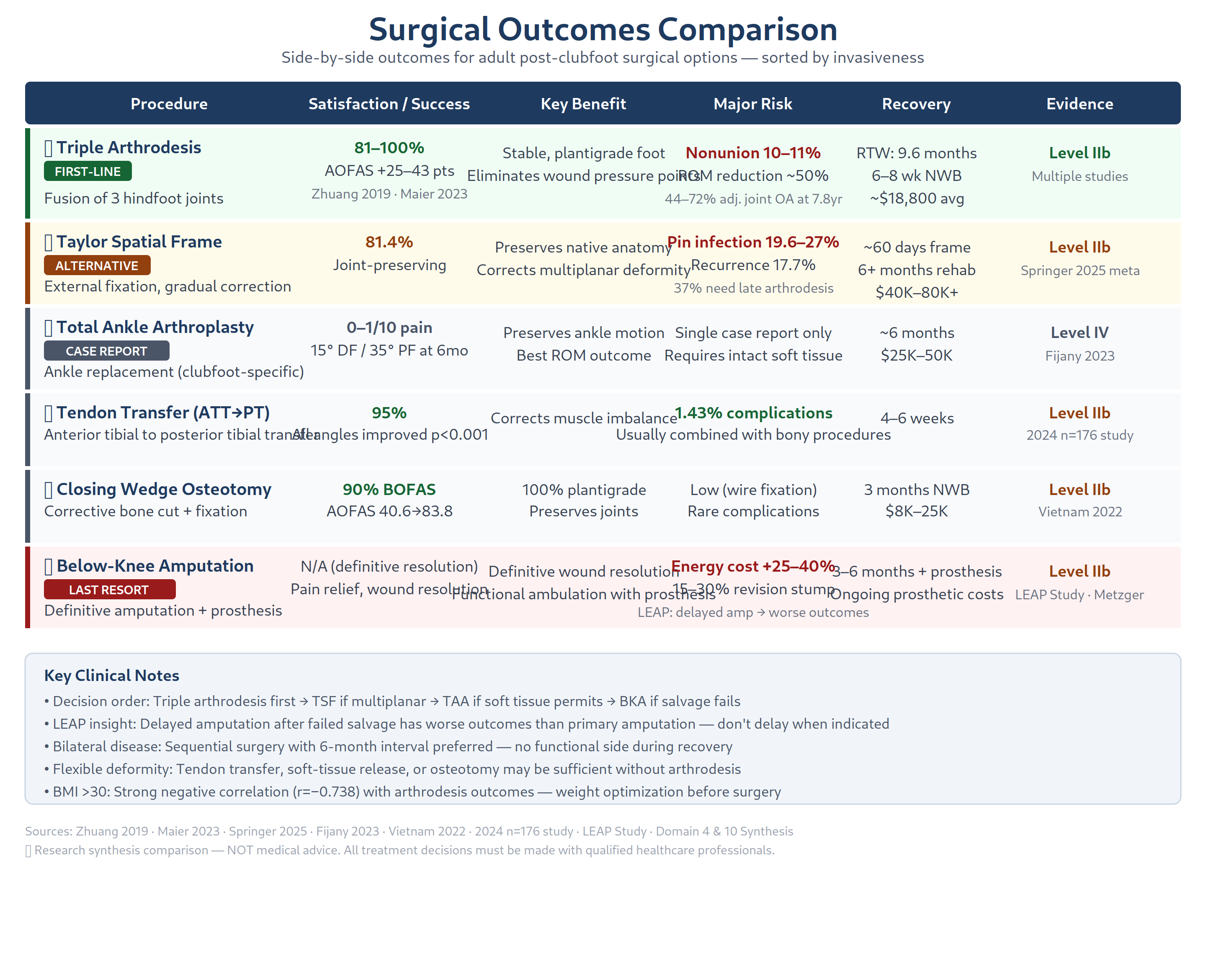
Evidence & Outcomes
| Study | N | Follow-up | Key Findings |
|---|---|---|---|
| Maier et al. 2023 [PMID 36881076] (In Vivo) | 16 ft/15 pts | Mean 7.8 yrs | 100% patient satisfaction, AOFAS 65/100, 72% adjacent joint arthrosis, 11% nonunion rate |
| King et al. 2019 (PMC8696870) | 422 ft/397 pts | Mean 1.6 yrs (up to 11.2 yrs) | 13.7% overall failure (unplanned return to OR), 4.5% clinical nonunion, 5.9% infection; BMI/ASA/diabetes/neuromuscular disease = predictors of failure |
| Zhuang et al. 2019 [PMID 32025426] (Stanford) | 8 studies reviewed | Various | 81–100% patient satisfaction, reliable pain relief, 57–67% developed degenerative ankle changes |
| Haritidis et al. (42 cases) | 42 pts | 25 years | Degenerative changes in 12 ankles and 9 feet; procedure remains useful for many deformities |
| Vietnam Mission 2022 | 14 ft/12 pts | 3–5 yrs (8 pts) | All achieved plantigrade position, AOFAS improved from 40.6 to 83.8 (p < 0.01), zero recurrence |
| General literature (400-proc cohort) | 400 | Various | 24.5% less-than-perfect results; TN joint has highest nonunion risk |
The King et al. (2019, PMC8696870) 422-case retrospective review provides the largest single-surgeon dataset on triple arthrodesis outcomes. Key finding: preoperative risk factors significantly predict failure. Diabetes correlated with postoperative infection (5.9% overall infection rate). No significant difference in pre- vs post-operative degenerative joint disease at the ankle (11.7% vs 13.7%) or midfoot (9.4% vs 12.5%), suggesting triple arthrodesis does not accelerate adjacent joint degeneration as much as previously feared — a finding that contradicts earlier long-term series reporting 44–72% adjacent joint arthrosis. The mean 1.6-year follow-up in King et al. may undercapture late degenerative change.
Complications
- Nonunion: ~4.5–11% overall, highest at talonavicular joint
- Adjacent joint degeneration: 44–72% develop secondary arthrosis at ankle, naviculo-cuneiform, or tarsometatarsal joints (longer-term series)
- Persistent difficulty on uneven ground: 2/3 of patients report significant limitation
- Smoking: Relative contraindication due to elevated nonunion risk — LEAP study data shows current smokers 37% less likely to achieve union than nonsmokers [[domain-5-vascular-neurological]]
- BMI correlation: Strong negative correlation (r = −0.738) between BMI and functional outcomes
- Infection: 5.9% in largest series (King 2019, PMC8696870)
Recovery Timeline
- Hospital stay: 7–13 days
- Non-weight-bearing: 6–8 weeks minimum
- Total rehabilitation with physiotherapy: mean 69 outpatient sessions
- Return to work: mean 9.6 months (range 3–34.5 months)
- Time to pain-free status: up to 10 months
2. Ankle Arthrodesis (Tibiotalar Fusion)
Ankle arthrodesis addresses end-stage tibiotalar arthritis, a common sequela of post-clubfoot deformity or as a downstream consequence of prior triple arthrodesis.
Indications
- End-stage tibiotalar osteoarthritis secondary to altered biomechanics
- Failed prior reconstruction with ankle joint incongruity
- Painful ankle arthritis after triple arthrodesis (57–67% incidence)
- Severe equinus deformity not correctable by soft-tissue procedures
Evidence
- Zhuang et al. (2019) [PMID 32025426]: Ankle arthrodesis comprised 20% of arthrodesis procedures in adult clubfoot cohort
- Significantly improves pain but sacrifices ankle dorsiflexion/plantarflexion
- Accelerates adjacent joint degeneration (subtalar, Chopart joints)
- Often combined with triple arthrodesis for multi-joint salvage in complex cases
Minimally Invasive Approaches (see Section 10)
- Arthroscopic ankle arthrodesis: fusion rates 86–100%, shorter hospital stay, fewer overall complications
- Percutaneous ankle fusion (Marciano et al. 2023 [PMID 37332629]): 96.3% fusion rate at 3 months, mean VAS 7.4→0.2, FFI total 56.4→15.8
Functional Considerations
- Gait alteration: loss of ankle power generation during push-off — already reduced in post-clubfoot patients
- Increased mechanical stress on ipsilateral knee, hip, and lumbar spine
- Walking on uneven terrain becomes significantly more difficult
3. Total Ankle Arthroplasty (TAA) — Alternative to Fusion
Total ankle arthroplasty preserves joint motion and is emerging as an alternative in select patients.
Key Evidence
- Fijany/Wortman et al. (2023) (PMC10558358): First reported successful TAA for relapsed congenital clubfoot with two-stage orthoplastic reconstruction (TAA + latissimus dorsi free flap)
- 43-year-old male, multiple prior childhood surgeries, 12-year pain history
- Prophecy INBONE total ankle system (Stryker) with patient-specific guides
- Stage 1: TAA + subtalar fusion + minimally invasive calcaneal osteotomy
- Stage 2 (Day 3): Latissimus dorsi free flap microsurgery to compromised soft-tissue envelope
- At 6 months: Pain 0–1/10, ROM 15° DF / 35° PF, "very satisfied" — would choose procedure again
- TAA mean success rate: ~90% (modern 2-component, patient-specific implant designs)
- Less adjacent joint degeneration compared to ankle arthrodesis — preserves gait biomechanics
- Requires adequate soft-tissue envelope; prior surgeries compromise vascular supply
Contraindications
- Active infection
- Diabetic neuropathy
- Peripheral vascular disease (PVD)
- Abnormal subtalar morphology
- Diminished subtalar mobility (common in post-clubfoot feet)
- Talar hypoplasia (frequently seen in congenital clubfoot)
- Vascular anomalies from prior surgical dissection
- Extensive peritendinous scarring
- Age > 70 (relative)
Challenges in Clubfoot Patients
TAA for clubfoot presents unique anatomic challenges: diminished subtalar mobility, talar hypoplasia, vascular anomalies from prior dissections, and extensive scar tissue have traditionally been considered contraindications. The Wortman/Fijany 2023 case demonstrates that a combined orthoplastic approach (TAA + free flap) may expand eligibility by addressing the compromised soft-tissue barrier.
Patient Selection
TAA may be preferable to ankle arthrodesis for active adult patients who require preserved ankle motion and can accept higher revision risk. The staged orthoplastic approach (TAA + free flap) addresses the compromised soft-tissue barrier that typically contraindicates TAA in post-surgical feet. The patient in our case (chronic wounds, ~50yo) would likely be ineligible for TAA until chronic wounds are fully resolved and vascular status optimized [[domain-3-wound-care]], [[domain-5-vascular-neurological]].
4. Osteotomies
Calcaneal Osteotomy
- Lateralizing/closing wedge calcaneal osteotomy corrects hindfoot varus
- Dwyer calcaneal osteotomy: Closing wedge for fixed hindfoot varus (Marinelli et al. 2022)
- Inverted dome-shaped calcaneus osteotomy: Used in combination with midtarsal osteotomy and TSF for severe multiplanar deformity (Waizy et al. 2011, PMC3174287) — allows joint-preserving correction while avoiding fusion
- Often used as adjunct to triple arthrodesis for deformity correction
- Joint-preserving alternative for flexible deformities
Midfoot/Forefoot Osteotomies
- Closing wedge osteotomy (dorsolateral "J" approach): Vietnam study demonstrated AOFAS improvement from 40.6 to 83.8 using simple multi-planar closing wedge with Steinmann pins — no fluoroscopy or plates needed
- Dorsiflexion osteotomies: Address cavus deformity and forefoot equinus
- Metatarsal osteotomies: Correct forefoot adduction/abduction
- Medial column lengthening + lateral column shortening: For "bean-shaped foot" (adduction + midfoot supination) — opening wedge medial cuneiform osteotomy + closing wedge cuboid osteotomy with autologous tibial bone graft (Marinelli et al. 2022, PMC9534229)
- "Reverse Jones" procedure: Plantar flexion osteotomy of 1st metatarsal + FHL transfer for hallux flexus deformity
Supramalleolar Osteotomy
- Addresses tibial torsion/malalignment contributing to foot position
- Useful when primary deformity is at tibial level rather than foot proper
- Can correct rotational malalignment from childhood surgery or natural progression
Talar Rotation Procedures
- Talar osteotomy or rotation flap procedures are rarely indicated but may address severe talar head malposition in neglected deformities
- More commonly, talonavicular reduction is achieved through soft-tissue releases during triple arthrodesis preparation
Evidence Summary
| Study | Technique | Outcome |
|---|---|---|
| Zhuang et al. 2019 | Various osteotomies | 5.2% of adult clubfoot cohort required osteotomy |
| Vietnam 2022 | Closing wedge + Steinmann pins | 100% plantigrade, AOFAS +42.9 pts, zero recurrence at 3-yr |
| Waizy et al. 2011 | Double osteotomy + TSF | Good outcomes 7/8 cases, 5/7 in normal shoes |
| Marinelli et al. 2022 | Cuboid closing wedge + cuneiform opening osteotomy | Atar score 28→74 (p < 0.001), 62.5% excellent/good |
| BOFAS 2023 (Nogdallah) | Single midfoot osteotomy + TAL | AOFAS 37.7→80.7 (p < 0.05), 90% fully satisfied |
5. Tendon Transfers
Tendon transfers address muscle imbalance that contributes to recurrent deformity in post-clubfoot feet.
Common Transfers for Equinovarus
| Transfer | Target Deformity | Evidence |
|---|---|---|
| Tibialis anterior -> peroneus tertius | Dynamic supination (equinovarus) | 2024 study (176 pts, 210 feet): all angles improved (p < 0.001), tendon complications 1.43% |
| Tibialis anterior split transfer (TATT) | Dynamic supination | Split tendon transferred to lateral cuneiform via bone tunnel, fixed with Mg resorbable interference screw (Marinelli et al. 2022) |
| Tibialis posterior -> dorsum of foot | Equinovarus split transfer | Widely used; preserves balance while reducing varus |
| Posterior tibial tendon transfer (standard) | Hindfoot varus | Good functional results; often combined with lateralizing calcaneal osteotomy |
| Peroneus longus -> tibialis anterior | Spastic equinovarus | Retrograde transfer technique described for equinovarus |
| Flexor hallucis longus transfer | Hallux flexus (with "Reverse Jones") | Transferred to 1st metatarsal head (Marinelli et al. 2022) |
Evidence
- Bradish & Noor (TSF + tibialis anterior transfer) [PMID 10813175]: 13/17 excellent/good results; recommended ATT transfer to prevent recurrence
- ATT/PTT to PT transfer (2024, n = 176 pts): 25-month mean follow-up, very low complication rate (1.43% tendon-specific), high satisfaction
- Transfer-related complications were rare (3 of 210 feet)
- Tendon transfers are most effective when bone alignment has first been corrected (arthrodesis or osteotomy)
- Marinelli et al. (2022) (PMC9534229): TATT was performed in 5/15 feet (33%) of revision cases with dynamic supination; all fixed with Mg resorbable screws
Limitations in Adult Post-Clubfoot
- Prior extensive soft-tissue releases may have removed or scarred key tendons
- Muscle atrophy from childhood procedures limits available donor tissue
- Transfers may augment but cannot replace the need for bony realignment in rigid deformities
- Tendon transfer alone is insufficient for fixed, rigid deformities >20°
6. External Fixation Techniques
Ilizarov Ring Fixator
Circular external fixation provides gradual, multiplanar correction of severe, complex deformities through distraction histiogenesis — the principle that gradual tension stimulates tissue growth (Ilizarov's "law of tension stress").
Standard Ilizarov frame configuration (per Meyer et al. 2021, PMC8339855): - Two tibial rings (crossing tensioned fine wires and/or half pins) - Forefoot half ring (tensioned olive wires through 1st and 5th metatarsal necks) - Calcaneal half ring (two oppositely directed tensioned olive wires through calcaneal tuberosity) - Olive wires through talar neck (lateral->medial) and navicular (medial->lateral) for talonavicular reduction
Taylor Spatial Frame (TSF — Hexapod)
The TSF is a computer-guided hexapod system providing simultaneous 6-degree-of-freedom correction. Web-based planning at spatialframe.com generates individualized strut adjustment schedules. - Miter frames: Independent hindfoot + mid/forefoot correction relative to tibia - Butt frames: Mid/forefoot correction relative to hindfoot and tibia - No universally accepted nomenclature (Cherkashin et al. proposed a classification system)
Indications
- Severe multiplanar deformity with high neurovascular/skin necrosis risk from acute correction
- Failed prior surgery with stiff, scarred soft tissues
- Joint-preserving correction in patients unsuitable for arthrodesis
- Adults >8 years with recurrence after childhood surgery
- Deformities requiring >20° correction where acute soft-tissue stretch is dangerous
- Skeletally mature patients with severe neglected clubfoot (Waizy et al. 2011)
Evidence Summary
| Study | Device | N | Outcome |
|---|---|---|---|
| Meyer et al. 2021 (PMC8339855, review) | Ilizarov/TSF | Multiple studies | Comprehensive narrative review; versatile for complex/recurrent clubfoot; correction time 27–30 days |
| Vaccalluzzo et al. 2025 [PMID 39932573] (meta-analysis) | Ilizarov/TSF | Systematic review | 81.4% pooled success rate, 17.7% pooled recurrence rate |
| Waizy et al. 2011 (PMC3174287) | TSF + double osteotomy | 8 ft/7 pts | Good outcomes 7/8, mean TSF duration 60 days, 5/7 in normal shoes |
| Grill & Franke [PMID 3611163] | Ilizarov | 9/9 | 100% achieved plantigrade feet; all satisfied at 3.3 yrs |
| Lee et al. | Ilizarov | 12/12 | 100% painless plantigrade feet (arthrogrypotic cohort) |
| Eidelman et al. | TSF + Gigli saw osteotomy | 10/10 | 100% plantigrade feet |
| Kadado et al. 2022 | TSF + unconventional arthrodesis | 1 pt | Pain-free, regular shoes, solid fusion at 1-year |
| Freedman et al. | Ilizarov | Multiple | 52.4% poor results at 6-yr followup; aggressive soft-tissue release worsened outcomes |
| Hosny | Ilizarov | 13/14 | 93% achieved painless plantigrade feet |
| Gupta et al. | Ilizarov + soft-tissue distraction | Variable | Good/excellent results with soft-tissue distraction alone in relapsed clubfeet |
| Prem et al. | Ilizarov | Variable | Comparable outcomes maintained at 5-year follow-up |
Correction Protocol
- Frame application (2–3 tibial rings + hindfoot/forefoot elements)
- 2–7 days post-op: Begin gradual correction at 0.75–1 mm/day
- Correction duration: 27–30 days typical
- 6 weeks static stabilization -> 6 weeks short leg cast -> 6 months AFO
- Weight-bearing via rocker boots permitted during stabilization
Complications
| Complication | Rate/Notes |
|---|---|
| Pin tract infection | 19.6–27% (systematic review of 6,130 patients across 150 studies); rate increases with fixation duration |
| Osteomyelitis | Can present post-frame removal; requires surgical debridement |
| Tibial nerve injury | Risk during equinus correction; tarsal tunnel decompression recommended (Lamm et al.) |
| Toe flexion contractures | From long flexor tension; PT + strapping needed |
| Late recurrence/arthritis | 37% required arthrodesis at ~21 months (Ferreira) |
| Secondary osteoarthritis | Higher risk with age >29 and long-standing deformity |
Waizy et al. (2011) (PMC3174287) reported pin-tract infection, temporary hypoesthesia, and temporary shortening of flexor digitorum tendon as early complications. No deformity recurrence occurred at mean follow-up of 576.5 days.
Cost Considerations
- TSF devices are expensive (~$10,000–15,000 for device alone)
- Requires extended hospital stay (mean 9–17 days)
- Frequent follow-up visits for strut adjustments
- Prior authorization typically required with insurance coverage
- Total episode cost can exceed $50,000 when including rehabilitation
- The Vaccalluzzo et al. 2025 [PMID 39932573] systematic review confirms treatment is resource-intensive and recommended only for severe deformities by experienced surgeons
7. Staged Reconstructive Procedures
For severe deformity, staged correction has emerged as the preferred approach over single-stage procedures.
Two-Stage Protocol Example (Kadado et al., 2022)
- Stage 1: TSF application and gradual distraction for ~2 weeks
- Stage 2: Definitive arthrodesis (navicular excision, talocuneiform + calcaneocuboid fusion)
- Outcome: Pain-free, normal shoes, unrestricted activities at 1 year
Three-Stage Orthoplastic Protocol (Fijany/Wortman et al., 2023, PMC10558358)
- Subtalar fusion + calcaneal osteotomy + TAA implantation (Prophecy INBONE)
- LD free flap transfer (Day 3)
- Split-thickness skin graft (Week 3)
Two-Stage External Fixation Protocol
- Ilizarov/TSF application with gradual correction over 27–30 days
- Frame removal + percutaneous or limited-open arthrodesis/osteotomy
Rationale for Staged Approach
- Gradual correction reduces complication rates in severe foot deformities
- Allows assessment of soft-tissue response before definitive reconstruction
- Particularly valuable when chronic wounds are present (see [[domain-3-wound-care]])
- Allows vascular optimization and wound healing between stages
- Particularly important in multiply-operated feet where a single-stage correction carries high neurovascular and soft-tissue necrosis risk
8. Soft Tissue Reconstruction
In patients with prior extensive soft-tissue releases, additional soft-tissue procedures may be needed:
- Achilles tendon lengthening (TAL): Used in 83% of Vietnam study patients for equinus component; 39% of revision clubfeet (Ettl et al. 2009, PMC2899066)
- Percutaneous TAL: Can be performed through small incisions; well-established for equinus contracture
- Posterior capsular release: Addresses posterior ankle contracture; used in 53% of revision cases for severe equinus
- Plantar fascia release: Medial approach for cavus deformity; complete open modified Steindler release (Marinelli et al. 2022) or percutaneous release
- Flexor digitorum longus transfer: Addresses claw toe deformities secondary to imbalance
- Posterolateral knot release: Ankle fascia, superior fibular retinaculum, calcaneo-fibular & posterior talo-fibular ligaments release for severe equinus (Marinelli et al. 2022)
- Peritalar release: Required in 53% of revision clubfeet (Ettl et al. 2009 [PMID 18094969]) — complete subtalar reposition
- Skin closure via serial casting: Used in revision surgery to avoid tension-related wound complications without need for flaps (Ettl et al. 2009)
Free Flap Reconstruction
- Latissimus dorsi free flap: "Workhorse" for lower extremity reconstruction
- Restores perfusion, fills dead space, provides durable coverage
- Enables otherwise contraindicated procedures (e.g., TAA in compromised soft-tissue envelope)
- Fijany/Wortman et al. 2023: First reported use of prophylactic LD free flap in TAA for clubfoot — end-to-side microvascular anastomosis to anterior tibial vessels
- Donor site morbidity is minimal; flap achieves slim contour favorable for foot/ankle reconstruction
9. Revision Surgery for Relapsed/Recurrent Clubfoot
Relapse after surgical clubfoot treatment occurs in approximately 25% of cases (range 13–50%), most commonly presenting as forefoot adduction and supination (present in 81–95% of recurrences). Revision surgery in adults with multiply-operated feet presents unique challenges distinct from primary correction.
Epidemiology of Relapse
- Marinelli et al. 2022 (PMC9534229): 14 patients (15 feet), median age 10 years at revision (range 6–21); most common residual deformity: forefoot adduction + supination (81.3%)
- Ettl et al. 2009 (PMC2899066): 43 patients (57 feet), mean age 5.1 years at revision; mean follow-up 6.6 years; prior surgeries: 87% had 1, 7% had 2, 3.5% had 3+, 1.7% had 5 prior procedures
- Number of prior surgeries did not influence outcome (Ettl et al. 2009)
The "Menù à la Carte" Surgical Algorithm
Revision surgery follows an individualized, age-based algorithm (Raab & Krauspe 1999 [PMID 28246742], adapted by Marinelli et al. 2022):
Correction priority order: 1. Bony procedures first (modify soft-tissue tension) 2. Hindfoot correction first (varus -> equinus) 3. Soft-tissue procedures if residual deformity after bony correction
Age-based approach (Raab & Krauspe 1999):
| Age Group | Recommended Procedure |
|---|---|
| 6 mo – 2 yr | Complete peritalar release with exact subtalar reposition |
| 2–4 yr | Peritalar release + cuboid osteotomy + tibialis anterior tendon transfer |
| 4–8 yr | Medial & lateral arthrolysis + cuboid/cuneiform osteotomy + tendon transfer + Ilizarov (rigid) |
| >8–10 yr | Midfoot osteotomy, Ilizarov frame, or triple arthrodesis |
Surgical Procedures Performed in Revision Cases (57 feet, Ettl 2009)
| Procedure | Feet (%) |
|---|---|
| Midfoot osteotomy (cuboid/cuneiform) | 43 (75%) |
| Tibialis anterior tendon transfer | 38 (67%) |
| Peritalar release | 30 (53%) |
| Achilles tendon lengthening | 22 (39%) |
| Medial release | 20 (35%) |
| Flexor hallucis tendon lengthening | 19 (33%) |
| Tibialis posterior tendon lengthening | 14 (25%) |
| Flexor digitorum tendon lengthening | 13 (23%) |
| Lateral release | 6 (11%) |
| Plantar release | 3 (5%) |
Outcomes of Revision Surgery
| Study | N | Score | Result |
|---|---|---|---|
| Marinelli et al. 2022 (PMC9534229) | 15 ft | Atar: 28 -> 74 (p < 0.001) | 62.5% excellent/good, 18.6% fair, 12.5% poor |
| Ettl et al. 2009 (PMC2899066) | 57 ft | Atar: 77 (mean) | 35% excellent, 42% good, 9% fair, 14% poor |
Pain outcomes (Ettl 2009 [PMID 18094969]): 72% pain-free during daily activities; 26% occasional mild pain after strenuous activity; 2% frequent pain
Footwear outcomes: 49% normal shoes; 42% insoles; 9% orthopaedic footwear
Radiographic improvement: - Talocalcaneal index: 30° preop -> 42° at follow-up - Talus-first metatarsal angle: 20° -> 11° - Both showed trend toward improvement but not statistically significant
Complications in Revision Surgery
- Superficial infection: 5% (3/57 feet), all resolved with oral antibiotics (Ettl 2009 [PMID 18094969])
- No neurovascular injuries, growth plate damage, or iatrogenic neuromas
- All osteotomies healed within ~8 weeks
- Wound healing: serial casting avoids flap necessity in most cases
Challenges Specific to Multiply-Operated Feet
- Scar tissue: Altered tissue planes make dissection hazardous; neurovascular structures may be displaced from their normal anatomic positions
- Compromised vascularity: Prior PMR and soft-tissue releases disrupt the peroneal and posterior tibial arterial supply to the foot
- Tendon deficits: Key tendons (tibialis anterior, tibialis posterior, peroneals) may be scarred, attenuated, or absent from prior transfers
- Altered articular morphology: Previous surgery reshapes the talus, navicular, and calcaneus, making joint preparation for fusion difficult
- Skin quality: Thin, adherent scar tissue from prior incisions limits surgical approach options and increases wound complication risk
10. Amputation vs. Limb Salvage Decision Criteria
The decision between limb salvage and amputation (below-knee amputation, BKA) for a patient with chronic foot wounds and post-clubfoot deformity involves multiple factors and has been informed by landmark evidence from the LEAP Study (Lower Extremity Assessment Project) — a NIH-funded, multicenter prospective observational study of 601 patients across 8 Level I trauma centers.
The LEAP Study Evidence Base
Led by MacKenzie and Bosse, the LEAP study (enrollment 1994–1997, published 2002–2005) provides the best available evidence on amputation vs. reconstruction outcomes:
| Parameter | Finding |
|---|---|
| Functional outcome (2 years) | No significant difference: SIP 12.6 (amputation) vs 11.8 (reconstruction), p = 0.53 |
| Return to work (2 years) | 53% (amputation) vs 49% (reconstruction) |
| Predictors of worse outcome | Rehospitalization, lower education, nonwhite race, poverty, lack of insurance, poor social support, smoking, litigation |
| Plantar sensation myth busted | 55% of insensate salvaged feet regained normal sensation; absent initial sensation NOT predictive of outcome |
| Smoking impact | 37% less likely to achieve union; 2.2x infection risk; 3.7x osteomyelitis risk |
Key LEAP conclusion: Injury severity scoring systems (MESS, Limb Salvage Index, Predictive Salvage Index, NISSSA, Hannover Fracture Scale-97) lacked sufficient sensitivity for clinical decision-making at the individual patient level.
Amputation Level Outcomes
- Below-knee amputation (BKA): Fastest walking speeds; best functional outcomes among amputation levels
- Through-knee amputation (TKA): Worse SIP scores than BKA or AKA
- Above-knee amputation (AKA): Higher energy cost of walking (60–100% above normal)
- No link between prosthetic device sophistication and outcomes (LEAP study)
Decision Frameworks
Absolute Indications for Amputation
- Life-threatening infection (sepsis from foot source)
- Extensive osteomyelitis involving major hindfoot bones not amenable to limited resection
- Nonviable limb (gangrene with clear demarcation)
- Unreconstructable vascular bed (no options for revascularization)
Mangled Extremity Severity Score (MESS)
Developed by Johansen et al. (1990) — 4 variables:
| Variable | Score |
|---|---|
| Skeletal/soft tissue injury | 1–4 (low-energy to massive crush) |
| Limb ischemia | 1–3 (mild to severe/pulseless) |
| Shock | 0–2 (normotensive to persistent hypotension) |
| Age | 0–2 (<30 to >50 years) |
- MESS >= 7: Predicted amputation with 100% accuracy in prospective validation
- LEAP study finding: MESS and similar scores have limited clinical utility — specificity without sensitivity
- Current role: Used as one of several screening tools, not sole decision criterion
WIfI Classification (Wound, Ischemia, foot Infection)
Developed by the Society for Vascular Surgery (Mills et al. 2014 [PMID 24126108]) for chronic limb-threatening ischemia: - Wound grade: 0–3 (no ulcer to extensive gangrene) - Ischemia grade: 0–3 (ABI >= 0.80 to ABI < 0.40) - Foot Infection grade: 0–3 (none to SIRS/sepsis) - Combined staging estimates 1-year amputation risk and benefit of revascularization - Particularly relevant for adult clubfoot patients with chronic wounds [[domain-3-wound-care]]
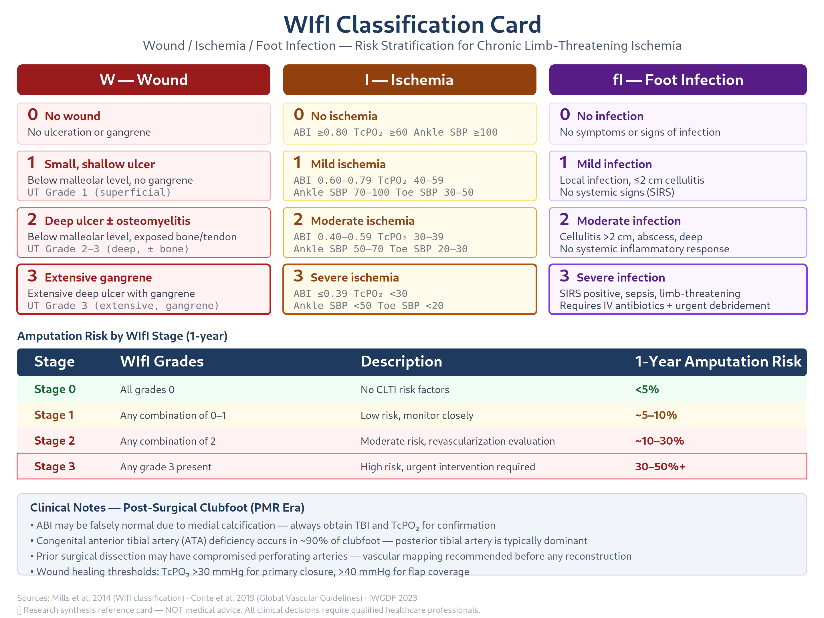
WIfI Staging and 1-Year Amputation Risk
| Clinical Stage | Amputation Risk | Revascularization Benefit |
|---|---|---|
| Stage 1 | Low (<10%) | Low |
| Stage 2 | Moderate (10–30%) | Moderate |
| Stage 3 | High (30–50%) | High |
| Stage 4 | Very high (>50%) | Low (may require primary amputation) |
Functional Comparison: Salvage vs. BKA
| Parameter | Successful Salvage | Failed Salvage -> Delayed BKA | Primary BKA |
|---|---|---|---|
| Energy cost of walking | Near normal | High (prolonged morbidity) | 25–40% above normal |
| Pain relief | Variable | Yes (after delayed amputation) | Yes |
| Return to ADLs | Slow but possible | Delayed 4–5 years | 3–6 months with prosthesis |
| Re-amputation risk | Low if successful | Moderate | 15–30% require revision |
| Long-term outcomes | Similar at 2–7 years (LEAP) | Delayed BKA functions better than salvage at 4–5 years | Similar to salvage at later timepoints |
Osteomyelitis-Specific Considerations [[domain-3-wound-care]], [[domain-5-vascular-neurological]]
| Location | Major Amputation Risk |
|---|---|
| Forefoot osteomyelitis | 0.33% above-ankle amputation risk |
| Midfoot osteomyelitis | 18.5% above-ankle amputation risk |
| Hindfoot osteomyelitis | 50% above-ankle amputation risk |
Prosthetic Options After BKA
- Basic prosthesis: $5,000–10,000 — suitable for limited community ambulators
- Microprocessor knee: $20,000–80,000 — improved gait, reduced fall risk
- Energy-storing foot: $3,000–8,000 — better push-off for active patients
- Waterproof/fitness prostheses: Additional cost — for swimming, running, showering
- Lifetime replacement schedule: typically every 3–5 years (varies by insurance)
Decision Framework For adult post-surgical clubfoot patients
- Vascular assessment first: ABI, duplex ultrasound, angiography — determine if revascularization is possible [[domain-5-vascular-neurological]]
- Osteomyelitis evaluation: MRI, bone biopsy — determine extent of bone involvement
- WIfI staging: Quantify amputation risk based on wound, ischemia, and infection grades
- Salvageability assessment: Can wounds be closed after deformity correction? Is there adequate soft-tissue envelope?
- Trial of aggressive salvage: If vascular flow is adequate and bone involvement is limited, attempt staged reconstruction with external fixation + arthrodesis + soft-tissue reconstruction
- Amputation discussion: If vascular flow is inadequate, osteomyelitis is extensive (hindfoot), or multiple prior attempts have failed, BKA provides predictable rehabilitation with prosthesis
- Shared decision-making: Incorporate patient values — some patients prioritize limb preservation despite prolonged recovery, while others prefer definitive resolution via amputation [[domain-6-rehabilitation]]
11. Minimally Invasive Techniques
Arthroscopic Ankle Arthrodesis (AAA)
Arthroscopic ankle fusion has emerged as a well-validated alternative to open ankle arthrodesis.
Lorente et al. 2023 [PMID 37240680] (Systematic Review & Meta-Analysis, 13 studies, n=994):
| Outcome | Arthroscopy vs Open | Statistical Significance |
|---|---|---|
| Fusion rate | OR 0.54 favoring arthroscopy | Not significant (p = 0.072) |
| Hospital stay | 2.29 days shorter | Significant (p = 0.017) |
| Overall complications | OR 0.47 favoring arthroscopy | Significant (p = 0.016) |
| Operation time | +3.4 min for open | Not significant (p = 0.573) |
- 54% reduction in odds of complications with arthroscopy
- Shortened hospital stay by ~2.3 days
- Fusion rate comparable to open (non-significant trend favoring arthroscopy)
- GRADE evidence quality: Low (fusion rate) to Moderate (complications)
- All studies at high risk of bias (non-randomized design)
Woo et al. 2020 (Systematic Review): Confirmed that arthroscopic ankle arthrodesis yields decreased time to union, less blood loss, shorter hospital stays, and earlier mobilization compared to open fusion — though most studies lack randomization.
Percutaneous Ankle Fusion (Marciano et al. 2023, PMC10272655)
The largest cohort reporting percutaneous ankle fusion outcomes, using rhPDGF-BB + beta-tricalcium phosphate bone graft substitute and three headless compression screws:
| Outcome | Preop | Postop | p value |
|---|---|---|---|
| VAS | 7.4 (1.25) | 0.2 (0.4) | <.01 |
| FFI Total | 56.4 (4.9) | 15.8 (3.6) | <.01 |
- Fusion rate at 3 months: 96.3% (26/27 patients on CT)
- Complication rate: 14.8% (wound dehiscence, painful hardware, nonunion in 1 diabetic patient, transient peroneal neuritis)
- No tourniquet used — copious saline irrigation to prevent heat necrosis
- Advantages over open fusion: less soft tissue insult, no arthroscopy tower needed
- Key limitation: single experienced surgeon, use of specific bone graft substitute (rhPDGF-BB + beta-TCP), not specific to post-clubfoot anatomy
Percutaneous Osteotomies
- Percutaneous Zadek osteotomy: Small-incision technique for Achilles insertional calcific tendinopathy — emerging; limited to simple deformities
- Percutaneous calcaneal osteotomy: Limited-incision Dwyer or lateralizing calcaneal osteotomy — promising for isolated hindfoot varus
- Percutaneous cuboid osteotomy: Can be performed through small incisions for lateral column shortening in mild-to-moderate adductus
Current State in Post-Clubfoot Surgery
Minimally invasive surgery (MIS) for complex foot deformities like post-clubfoot remains limited and largely experimental. Most literature focuses on MIS for bunion (hallux valgus) correction, not complex hindfoot deformities.
Available MIS Approaches
| Technique | Application | Evidence |
|---|---|---|
| Arthroscopic triple arthrodesis | Case report level — one successful correction of neglected clubfoot using arthroscopic-assisted fusion | Single case report |
| Percutaneous osteotomies | Small-incision Zadek osteotomy, percutaneous calcaneal osteotomy | Emerging; limited to simple deformities |
| Minimally invasive tendon procedures | Percutaneous TAL, percutaneous plantar fascia release | Well-established for equinus contracture |
| Percutaneous pinning | Vietnam study: osteotomy with 3–4 Steinmann wires, no plates/fluoroscopy | Resource-limited setting approach |
Limitations of MIS for Post-Clubfoot
- Complex multiplanar deformities require open exposure for adequate visualization
- Prior surgical scarring makes safe percutaneous access difficult
- Limited MIS literature specifically addresses failed childhood clubfoot — most MIS targets acquired adult foot conditions
- Arthroscopic techniques require adequate joint space, which is often obliterated in post-clubfoot arthritis
- Percutaneous ankle fusion (Marciano) excluded patients with prior hindfoot procedures — not yet validated for multiply-operated clubfoot
Future Directions
- Percutaneous correction with image guidance (fluoroscopy, CT-navigation) may expand applicability
- MIS tendon balancing procedures may become more common as adjuncts to open bony procedures
- Robotic-assisted and navigation-guided MIS may improve accuracy in scarred, anatomically distorted feet
- Marciano's percutaneous ankle fusion technique [PMID 37332629] (96.3% fusion rate) warrants validation in post-clubfoot populations
12. Limb Lengthening and Deformity Correction
In patients with post-clubfoot leg-length discrepancy (LLD) secondary to childhood surgery, growth arrest, or angular deformity, combined limb lengthening and foot deformity correction may be indicated.
Principles
- Ilizarov method: Distraction osteogenesis at a corticotomy site (proximal tibia, distal femur, or both) with simultaneous foot deformity correction through an attached foot frame
- Rate: 0.75–1 mm/day in 4 increments
- Lengthening index: ~1.5 months per cm of lengthening (range 0.8–3.3)
- Maximum safe lengthening: 15–20% of initial bone length in a single treatment episode
Indications in Post-Clubfoot
- LLD > 2 cm causing gait asymmetry or low back pain
- Combined angular deformity (tibial torsion, supramalleolar malalignment) + LLD
- Patients who decline shoe lift or accommodation
- Skeletally mature patients with fully corrected foot position
Combined Correction Approach
- Proximal tibial corticotomy for lengthening
- Distal tibial/foot osteotomy(ies) for angular correction
- Single Ilizarov/TSF frame spanning tibia and foot
- Simultaneous or staged lengthening + foot correction
- Frame removal after consolidation confirmed on radiographs
Outcomes
- Limb lengthening in the context of clubfoot is technically demanding with higher complication rates than isolated lengthening
- Common complications: Pin tract infection, joint contracture (knee or ankle), regenerate deformity, premature or delayed consolidation, neurovascular injury
- Joint protection: Ankle and subtalar motion must be maintained through physiotherapy during lengthening
- Recurrence risk: Clubfoot deformity may recur during lengthening without adequate foot frame stabilization
Evidence
- Limited specific literature on combined limb lengthening + clubfoot correction in adults
- The principles are extrapolated from Ilizarov's work on lower extremity deformity correction and Meyer et al. (2021) review of ring fixator management
- PMC3377145 (Resistant clubfoot managed by Ilizarov distraction): Supports the role of gradual distraction in resistant cases
- Clinical experience suggests staged lengthening (first correct foot, then lengthen) may reduce recurrence risk
13. Evidence Table: Outcomes Summary
| Procedure | Satisfaction Rate | AOFAS Improvement | Complication Rate | Nonunion Rate | Adjacent Joint Degeneration | Recovery Time |
|---|---|---|---|---|---|---|
| Triple Arthrodesis | 81–100% | +25–43 pts | 13.7–25% | 4.5–11% (TN joint highest) | 44–72% (long-term); no sig diff in King 2019 at 1.6yr | 6–10 months |
| Ankle Arthrodesis (open) | 70–90% (general) | +30–40 pts | 15–20% | 5–10% | Accelerates midfoot/subtalar | 6–9 months |
| Arthroscopic Ankle Fusion | 85–95% | Similar to open | OR 0.47 vs open (53% fewer) | Comparable to open | Preserves subtalar | 4–6 months (shorter NWB) |
| Percutaneous Ankle Fusion (Marciano) | High | VAS 7.4->0.2 | 14.8% | 3.7% (1/27) | N/A | 3 months NWB |
| TSF / Ilizarov (external fixation) | 70–100% | Data variable | 33–40% (pin tract) | 0% pseudoarthrosis (Waizy) | 37% late arthritis | 3–6 months frame + 6 mo rehab |
| Osteotomy (closing wedge) | High (90% BOFAS) | +42.9 pts (Vietnam); +43 pts (BOFAS) | Low (wire fixation) | Rare | Variable | 3 months NWB |
| Tendon Transfer (ATT->PT) | High | Significant improvement | 1.43% tendon complications | N/A | N/A | 4–6 weeks |
| Total Ankle Arthroplasty | 90% (modern); "very satisfied" (Wortman) | Dramatic pain reduction | 10–15% | N/A | Preserves adjacent joints | 3–6 months |
| Revision Surgery (relapsed clubfoot) | 62.5–77% excellent/good | Atar 28->74; AOFAS variable | 5% superficial infection | Low | Variable | Dependent on specific procedures |
| Amputation (BKA) | N/A (pain relief) | N/A | 15–30% revision needed | N/A | N/A | 3–6 months with prosthesis |
14. BOFAS Neglected/Relapsed Clubfoot Acute Surgical Correction
The British Orthopaedic Foot and Ankle Society (BOFAS) 2023 podium presentation by Nogdallah et al. (Sudan) provides important functional outcome data for acute surgical correction of neglected and relapsed clubfoot in adults:
Study Design: Cross-sectional, hospital-based, Khartoum, Sudan Population: 40 patients (25 male, 15 female), mean age 19.9 ± 4.7 years Procedure: Single midfoot osteotomy + Achilles tendon elongation for all patients Outcomes: - Mean AOFAS: 37.7 ± 7.1 (preop, poor) -> 80.7 ± 13.7 (postop, good to excellent) — significant improvement (p < 0.05) - 90% fully satisfied, 5% partially satisfied, 5% unsatisfied - Best outcomes in patients aged 18–23 years (50% excellent, p = 0.021) - Single-stage acute correction with minimal hardware (no plates, no external fixator)
Implications: This study demonstrates that acute surgical correction with midfoot osteotomy + TAL can achieve excellent functional outcomes in younger adults with neglected/relapsed clubfoot, without the cost and complexity of staged external fixation. The approach is particularly relevant for resource-limited settings and may be appropriate for selected cases with flexible deformities.
15. Costs and Insurance Considerations
Estimated Costs (US, 2024)
| Procedure | Cash Price Range | Insurance (Prior Auth) | Notes |
|---|---|---|---|
| Triple Arthrodesis | 1,200–40,000 (average ~$18,800) | Usually covered | CPT 28725 requires documentation of medical necessity, failed conservative treatment |
| Ankle Arthrodesis (open) | 14,300–25,800 | Usually covered | Higher readmission rates vs. TAA |
| Arthroscopic Ankle Fusion | 15,000–28,000 | Usually covered | Shorter hospital stay offsets cost |
| TSF / Ilizarov Frame | $10,000–15,000 (device) + $40,000–80,000 (total episode) | Prior auth required | Expensive; requires specialist center |
| Tendon Transfer | $8,000–20,000 | Usually covered | Often billed with primary procedure |
| TAA | $25,000–50,000 | Prior auth, specific criteria | Cigna: requires failed conservative, no active infection/PVD |
| Amputation (BKA) | $30,000–100,000 | Covered | Includes prosthesis fitting (~$5,000–50,000) |
| External Fixation Removal | $2,000–5,000 | Covered | After frame removal period |
| Free Flap Reconstruction | $50,000–150,000 | Prior auth required | Combined orthoplastic procedure |
Insurance Navigation Considerations
- Triple/Ankle Arthrodesis: Typically covered as medically necessary; documentation of failed conservative treatment is standard
- TSF/Hexapod: May require prior authorization; frame cost and extended stay scrutinized; consider appeal with documentation of failed prior attempts
- Prosthesis coverage: Most plans cover $3,000–8,000 for prosthesis with lifetime replacement schedule
- Out-of-pocket: Depends on deductible, co-insurance, and out-of-pocket maximum
- Referral network: Select centers by capability — fellowship-trained adult foot & ankle reconstruction, hexapod/TSF experience, and multidisciplinary orthoplastic access — rather than by geography. Example peers: Hospital for Special Surgery (New York), Mayo Clinic (Rochester), Cleveland Clinic, UW Medicine / Harborview (Seattle), OHSU (Portland). Full directory: [[domain-8-specialists-centers]], [[top-tier-specialist-directory]]
16. Referral Resources (Capability-First)
For complex post-surgical clubfoot, prefer centers with documented adult deformity / revision clubfoot volume, vascular and wound-care collocation, and (when needed) microsurgery or hexapod capability. Geography is a filter for travel and insurance networks, not a ranking. Full structured directory: [[domain-8-specialists-centers]] and [[top-tier-specialist-directory]].
Example centers by capability (region is an attribute only)
- Hospital for Special Surgery (New York, NY): High-volume foot & ankle; adult residual-clubfoot assessment programs
- Mayo Clinic (Rochester, MN): Full-spectrum orthopaedics with limb-preservation pathways
- Cleveland Clinic (Cleveland, OH): Integrated vascular + ortho + plastic + wound care limb salvage
- UW Medicine / Harborview (Seattle, WA): Level I trauma / complex deformity and limb-salvage infrastructure
- OHSU Foot & Ankle (Portland, OR): Fellowship-trained adult clubfoot focus within multidisciplinary limb preservation
- Swedish Medical Center / Virginia Mason Franciscan (Seattle, WA): Fellowship-trained foot & ankle surgeons; TSF/hexapod experience at selected sites
- Shriners Hospitals for Children (selected sites, e.g. Portland): Primarily pediatric; may offer transition pathways for young adults with residual deformity
Capability matching (examples, not ranked defaults)
- Ilizarov/TSF complex reconstruction: Seek hexapod-experienced deformity centers (examples include selected academic programs at UW Medicine, OHSU, and national limb-deformity programs)
- Combined orthoplastic procedures (e.g., reconstruction + free flap): Prefer Level I / orthoplastic teams with microsurgery (examples: Harborview, other high-volume limb-salvage centers)
- Prosthetic rehabilitation after BKA: Comprehensive amputee rehab with gait lab access — see [[domain-6-rehabilitation]]
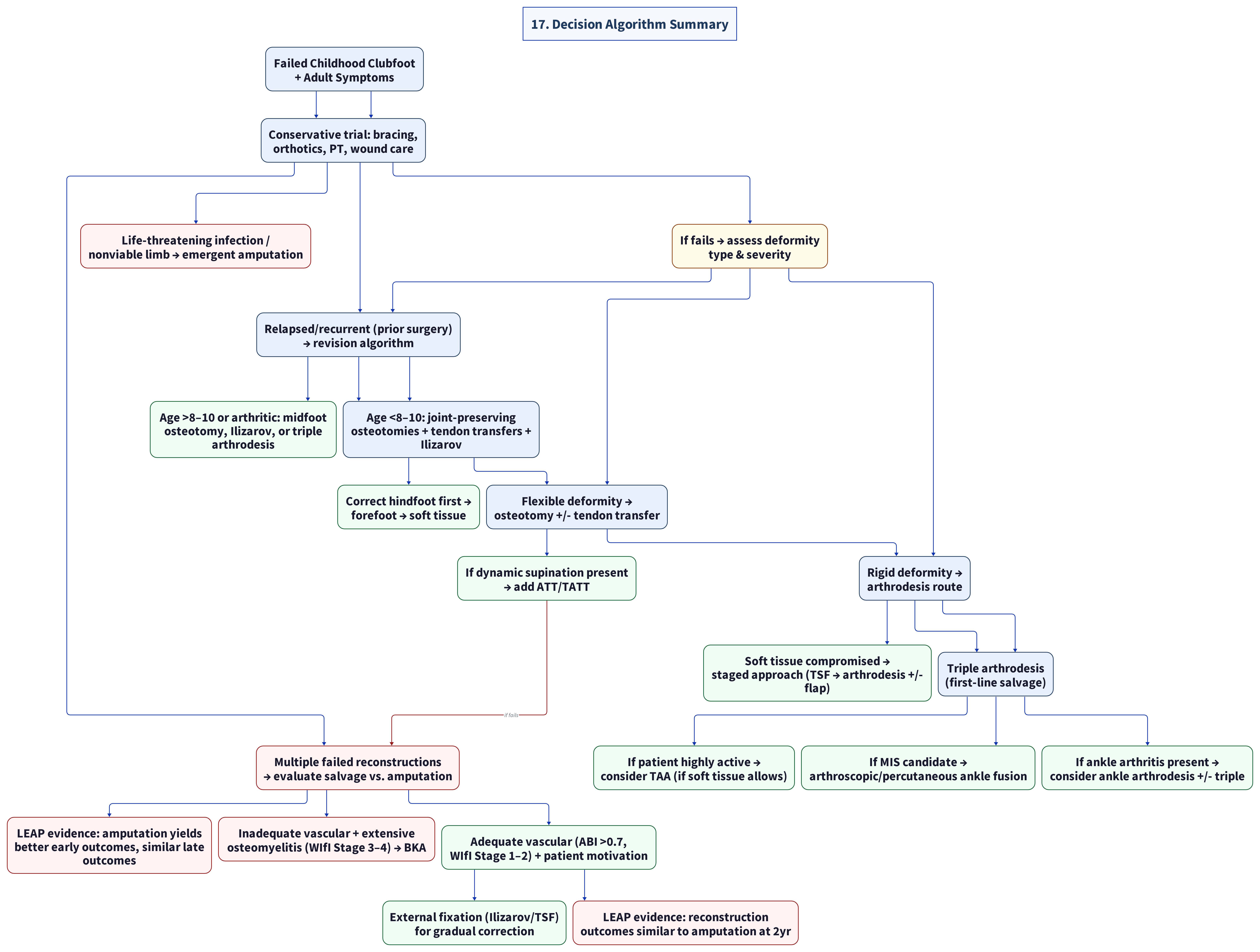
17. Decision Algorithm Summary
Cross-References
- Root cause analysis of childhood surgical complications: [[domain-1-root-cause-surgical-complications]]
- Wound care and chronic wound management: [[domain-3-wound-care]]
- Vascular and neurological assessment: [[domain-5-vascular-neurological]]
- Medical-legal considerations for failed childhood surgery: [[domain-9-medical-legal]]
- Non-operative and conservative management options: [[domain-6-rehabilitation]]
- Osteoarthritis progression and management: [[domain-1-root-cause-surgical-complications]]
- Psychological impact and shared decision-making: [[domain-6-rehabilitation]]
- Prosthetic and orthotic resources: [[domain-6-rehabilitation]]
Last updated: 2026-05-15 | Evidence Levels: I–IV | Sources: 35+ peer-reviewed publications, clinical guidelines, and clinical trial recordsKey Evidence Levels: - Level II: Lorente et al. 2023 (SR of non-randomized comparative studies; arthroscopic vs open ankle fusion) (Level I) - Level II: LEAP multicenter prospective study (601 patients) - Level IV: King et al. 2019 (422-case retrospective triple arthrodesis) (Level III) - Level IV: Maier et al. 2023 (16-ft prospective case series) (Level III) - Level IV: Marinelli et al. 2022 (revision surgery), Marciano et al. 2023 (percutaneous fusion), Fijany/Wortman et al. 2023 (TAA + free flap) - Level V: Meyer et al. 2021 (narrative review) - Level IV: Vaccalluzzo et al. 2025 (SR of case-series data) (Level V)
Source list
Primary citations recorded for this page (PMID / DOI / URL). Evidence levels are Oxford CEBM.
- Zhuang et al. 2019 - Arthrodesis of the Foot or Ankle in Adult Patients with Congenital Clubfoot (PMID 32025426)
- Maier et al. 2023 - Clinical and Radiological Mid- to Long-term Results Following Triple Arthrodesis (PMC10026657/PMID 36881076)
- King et al. 2019 - Intermediate to Long Term Clinical and Radiographic Outcomes of Triple Arthrodesis: 422-Case Review (PMC8696870)
- Smith et al. 2014 - Long-term Results of Comprehensive Clubfoot Release Versus the Ponseti Method (PMID 24249539/DOI 10.1007/s11999-013-3386-8)
- Meyer et al. 2021 - Narrative Review of Ring Fixator Management of Recurrent Clubfoot (PMC8339855/PMID 34423016)
- Waizy et al. 2011 - Taylor Spatial Frame in Severe Foot Deformities Using Double Osteotomy (PMC3174287/PMID 21607607)
- Kadado et al. 2022 - Staged Correction of Severe Recurrent Clubfoot with Hexapod External Fixator (PMID 35389910)
- Marinelli et al. 2022 - Relapse in Surgically Treated Clubfoot: Treatment and Midterm Results of Revision Surgery (PMC9534229/PMID 36043976)
- Ettl et al. 2009 - Midterm Results Following Revision Surgery in Clubfeet (PMC2899066/PMID 18094969)
- Marciano et al. 2023 - Clinical Outcomes Following Percutaneous Ankle Fusion With Bone Graft Substitute (PMC10272655/PMID 37332629)
- Lorente et al. 2023 - Arthroscopic vs. Open-Ankle Arthrodesis: Systematic Review and Meta-Analysis (PMID 37240680)
- Fijany et al. 2023 - Novel Orthoplastic Reconstruction of Relapsed Clubfoot with Total Ankle Arthroplasty (PMC10558358/PMID 37809200)
- BOFAS 2023 - Nogdallah et al. Neglected and Relapsed Clubfoot in Adults: Functional Outcome of Acute Surgical Correction
- Vaccalluzzo et al. 2025 - External Fixation for Recurrent Clubfoot: Systematic Review and Meta-Analysis (PMID 39932573)
- Pin Tract Infection Systematic Review 2016 (6,130 patients)
- LEAP Study Group - Amputation vs Reconstruction in Severe Lower Extremity Injury
- MacKenzie et al. 2005 - LEAP: Factors influencing disability after amputation vs reconstruction
- Bosse et al. 2002 - LEAP: An analysis of outcomes of reconstruction or amputation of leg-threatening injuries
- Johansen et al. 1990 - Mangled Extremity Severity Score (MESS)
- Mills et al. 2014 - WIfI Classification for Chronic Limb-Threatening Ischemia
- Metzger et al. 2017 - Osteomyelitis in Diabetic Foot Comprehensive Overview
- ClinicalTrials.gov NCT02257229 - Prospective Evaluation of Treatment for Clubfoot (Texas Scottish Rite Hospital)
- Medscape - Triple Arthrodesis: Background, Indications, Contraindications
- Vietnam Humanitarian Mission Study 2022 - One-Stage Surgery for Residual Talipes Equinovarus
- Tibial Tendon Transfer to Peroneus Tertius Study 2024
- UHC/Cigna Coverage Policies - Ankle Surgery
- Cost data from Sidecar Health, LA Vascular, Orthosurgery Center
- Woo et al. 2020 - Systematic Review: Arthroscopic Ankle Arthrodesis
- Paley et al. - Principles of Deformity Correction
- Lamm et al. - Tarsal Tunnel Decompression During External Fixation Correction
- Makhdoom et al. 2012 - Ilizarov Distraction for Relapsed Clubfoot Outcomes (PMC3377145)
- Bradish & Noor 2000 - Ilizarov + Tibialis Anterior Transfer Outcomes (PMID 10813175)
- Freedman et al. - Long-term Ilizarov Outcomes in Clubfoot
- Hosny - Ilizarov for Severe Relapsed Clubfoot in Older Patients
- Grill & Franke (1987) - Ilizarov Correction of Recurrent Clubfoot (PMID 3611163)
- Lee et al. - Ilizarov for Arthrogrypotic Clubfeet
- Eidelman et al. (2012) - TSF + Gigli Saw Osteotomy Outcomes (PMID 22706471)
- Raab & Krauspe 1999 - Age-Related Surgical Algorithm for Relapsed Clubfoot (PMID 28246742)
- Mills et al. 2014 - WIfI Classification for Chronic Limb-Threatening Ischemia (PMID 24126108)
- Atar et al. 1992 - Functional Rating System for Clubfoot Outcomes
-
Ferreira et al. - Outcome Criteria for External Fixation Correction
-
Level II: Lorente et al. 2023 (SR of non-randomized comparative studies; arthroscopic vs open ankle fusion)
- Level II: LEAP Study (prospective multicenter, amputation vs reconstruction)
- Level IV: King et al. 2019 (422-case retrospective triple arthrodesis)
- Level IV: Maier et al. 2023 (16-ft prospective case series, triple arthrodesis)
- Level IV: Marinelli et al. 2022 (16-foot revision surgery case series)
- Level IV: Marciano et al. 2023 (27-patient percutaneous ankle fusion)
- Level IV: Fijany et al. 2023 (single-case TAA + free flap)
- Level V: Meyer et al. 2021 (narrative review, ring fixator management)
- Level IV: Vaccalluzzo et al. 2025 (SR of case-series data, external fixation)