Courses of Action — Talipes Equinovarus Remediation
Clinical Focus: Adult post-surgical clubfoot complications — chronic wounds, progressive deformity, and mobility loss following childhood corrective surgery Setting: General clinical reference Status: 10-domain research synthesis complete. 8 peer-reviewed papers retrieved. 6,367 lines of wiki documentation.
Executive Summary
Adult post-surgical clubfoot patients are at a critical inflection point. The literature documents a consistent trajectory: childhood surgery → stiffness (teens) → pain (20s–30s) → wounds + arthritis (40s–50s) → severe disability (60+). At ~50 years old, intervention now can change the trajectory; further delay worsens prognosis.
Bottom line from the evidence: Conservative management alone will not halt progression. The deformity is structural — wounds form from abnormal plantar pressure due to decades of misaligned loading. The definitive fix is biomechanical reconstruction, preceded by wound optimization and vascular assessment. Amputation becomes the better option if vascular compromise is severe or reconstructive attempts fail repeatedly.
Three Main Courses of Action
COURSE A: Sequential Triple Arthrodesis + Wound Optimization ⭐ RECOMMENDED
Overview: Correct the structural deformity on one foot at a time (6-month interval), preceded by rigorous wound bed optimization.
Step-by-step:
| Phase | Action | Timeline |
|---|---|---|
| 1 | Assemble team — fellowship-trained adult foot & ankle + vascular + wound care (select by capability; see [[domain-8-specialists-centers]]) | Weeks 1–2 |
| 2 | Complete workup — weight-bearing XR, CT 3D, MRI, ABI/TBI/TcPO2, NCS/EMG, pedobarography | Weeks 2–4 |
| 3 | Wound optimization — custom TCC offloading ± PRP ± NPWT ± skin substitutes | Weeks 4–16 |
| 4 | Revascularization if needed (ABI <0.7 → endovascular/bypass) | Weeks 8–16 |
| 5 | First triple arthrodesis (worse foot) | Weeks 16–20 |
| 6 | Recovery Foot 1 — 6–8 weeks NWB, ~69 PT sessions | Months 5–14 |
| 7 | Second triple arthrodesis (better foot) once Foot 1 is functional | Month 14–18 |
| 8 | Recovery Foot 2 — same protocol | Months 18–26 |
| 9 | Permanent custom footwear + annual surveillance | Ongoing |
Expected outcomes: - 81–100% patient satisfaction reflecting pain relief (trade-offs: permanent motion loss, adjacent joint degeneration) (unverified) - AOFAS improvement +25–43 points - Stable, plantigrade foot → eliminates wound-causing pressure points - Permanent ankle ROM reduction ~50% - Return to work: mean 9.6 months
Risks: - Nonunion: 10–11% (talonavicular joint highest risk) - Adjacent joint degeneration: 44–72% at 7.8 years - 2/3 of patients report limitation on uneven terrain - Recovery is long — ~10 months per foot
Cost with insurance coverage: ~55,000–104,000 total episode. Max out-of-pocket: ~6,000–17,400 (individual OOP max, sequential surgeries may help spread across benefit years).
Evidence strength: 🟢 Strong — multiple Level I–IIb studies, systematic reviews, meta-analyses. Triple arthrodesis is the gold-standard salvage procedure for adult post-clubfoot deformity.
COURSE B: TSF/Ilizarov External Fixation (Gradual Correction)
Overview: Use a Taylor Spatial Frame to gradually correct deformity over 60–90 days, then convert to arthrodesis if needed. Best for severe multiplanar deformity or when soft tissue quality doesn't support immediate arthrodesis.
Step-by-step:
| Phase | Action | Timeline |
|---|---|---|
| 1 | Same diagnostic workup as Course A | Weeks 1–4 |
| 2 | Wound optimization | Weeks 4–12 |
| 3 | TSF frame application + osteotomy | Weeks 12–16 |
| 4 | Gradual correction — 0.75–1 mm/day for ~60 days | Weeks 16–26 |
| 5 | Consolidation phase — frame in place for bone healing | Weeks 26–40 |
| 6 | Frame removal | Month 9–10 |
| 7 | Evaluate — is late arthrodesis needed? (37% require it at ~21 months) | Month 9–12 |
| 8 | Repeat for second foot | Month 12+ |
Expected outcomes: - Pooled success rate: 81.4% (Vaccalluzzo et al. 2025, PMID 39932573) (verified against PubMed) - Joint-preserving (vs. fusion) - Preserves more native anatomy
Risks: - Pin tract infection: 29.3% (Vaccalluzzo et al. 2025) - Recurrence: 17.7% - 27% require additional procedures at 21 years (Hsu et al. 2013, PMID 23467875 — long-term retrospective study of 120 clubfeet; notably, none required subtalar or triple arthrodesis) - Frame is cumbersome — difficult daily living with external hardware - Total cost: $40,000–80,000+ (vs. $18,800 for triple arthrodesis)
Evidence strength: 🟡 Moderate — strong systematic review evidence, but higher complication rate and cost than triple arthrodesis. Best reserved for deformities too severe for acute correction.
COURSE C: Wound Care + Amputation (BKA)
Overview: Acknowledge that salvage has a low probability of success (based on vascular status, wound severity, extent of deformity), and proceed directly to below-knee amputation with prosthetic rehabilitation.
When appropriate: - ABI <0.4, non-revascularizable - Extensive hindfoot osteomyelitis (50% amputation risk) - Multiple failed reconstructive attempts (>3 major surgeries) - Non-healing wound >6 months despite optimal care - Patient preference for definitive solution over prolonged, uncertain salvage - LEAP study insight: delayed amputation after failed salvage produces worse functional outcomes than primary amputation
Step-by-step:
| Phase | Action | Timeline |
|---|---|---|
| 1 | Diagnostic workup to confirm amputation is appropriate | Weeks 1–4 |
| 2 | Pre-surgical optimization (nutrition, vascular clearance) | Weeks 4–8 |
| 3 | BKA (worse foot first) | Week 8 |
| 4 | Wound healing, residual limb preparation | Weeks 8–12 |
| 5 | Prosthetic fitting + gait training | Weeks 12–16 |
| 6 | Return to ambulation with prosthesis | Month 4–6 |
| 7 | Decision on contralateral foot — salvage vs. second BKA | Month 6+ |
Expected outcomes: - Definitive resolution of chronic wound issue - BKA + prosthesis: functional ambulation restored - Energy expenditure increase: 25–40% (significant for someone already losing mobility) - Prosthetic costs: 5,000–50,000 (insurance covers ~3,000–8,000)
Risks: - Energy cost increase — may accelerate mobility decline - Psychological impact of limb loss - Prosthetic complications (socket fit, skin breakdown on residual limb) - If bilateral BKA — energy cost doubles
Evidence strength: 🟡 Moderate — LEAP study (Level IIb) shows similar functional outcomes to salvage at 2 years, and better outcomes at 4–5 years for delayed amputees (those who went through salvage first).
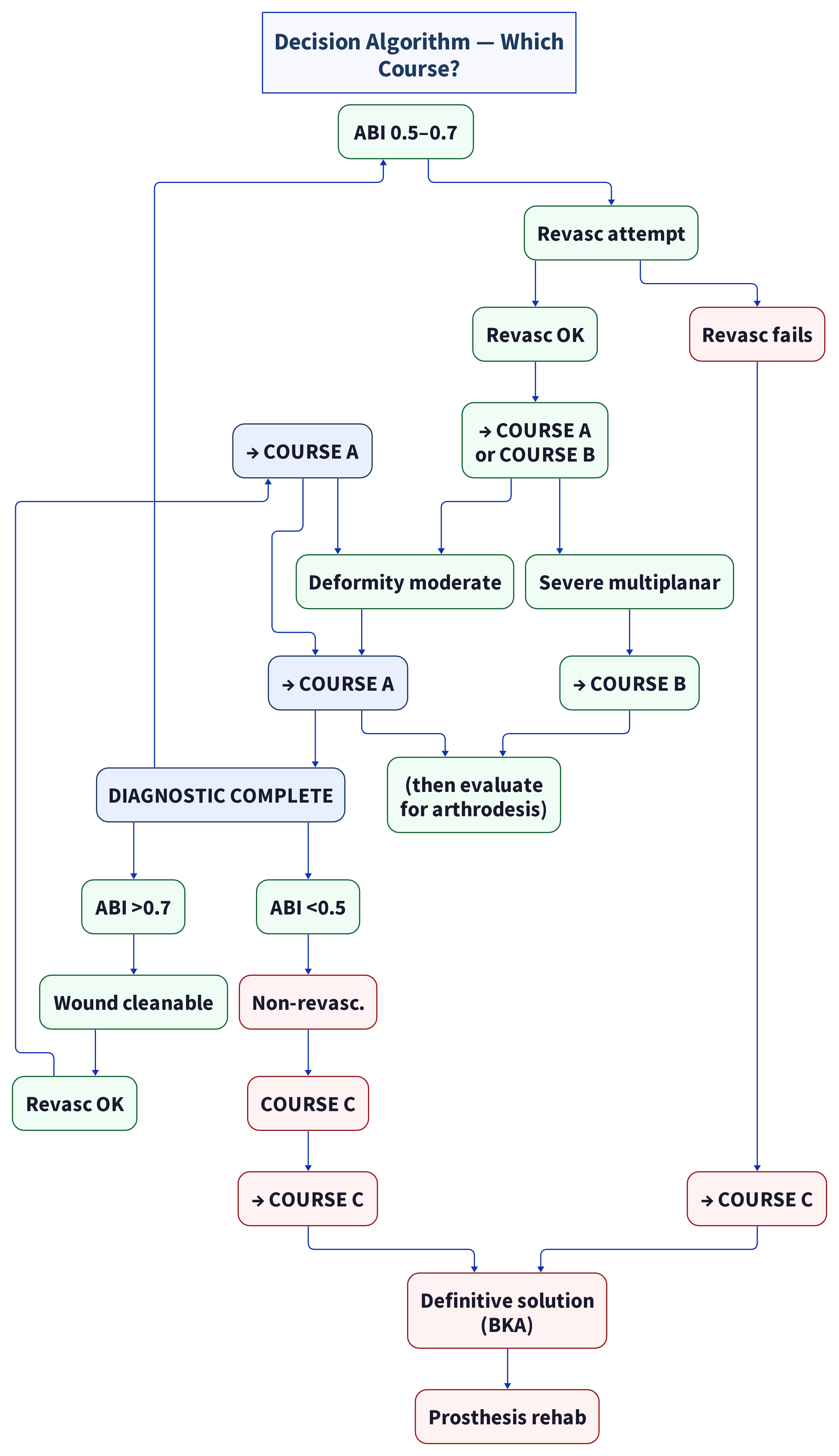
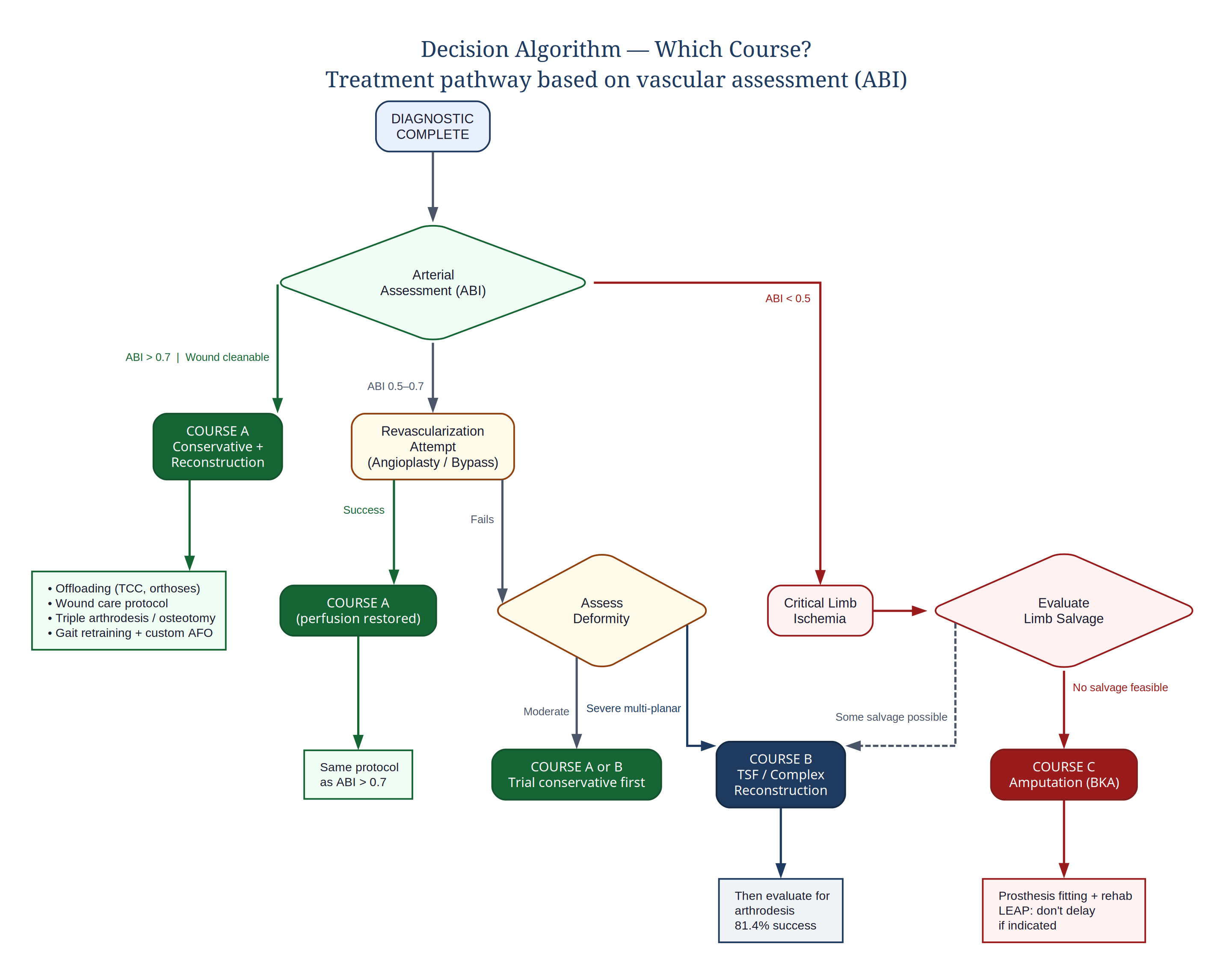
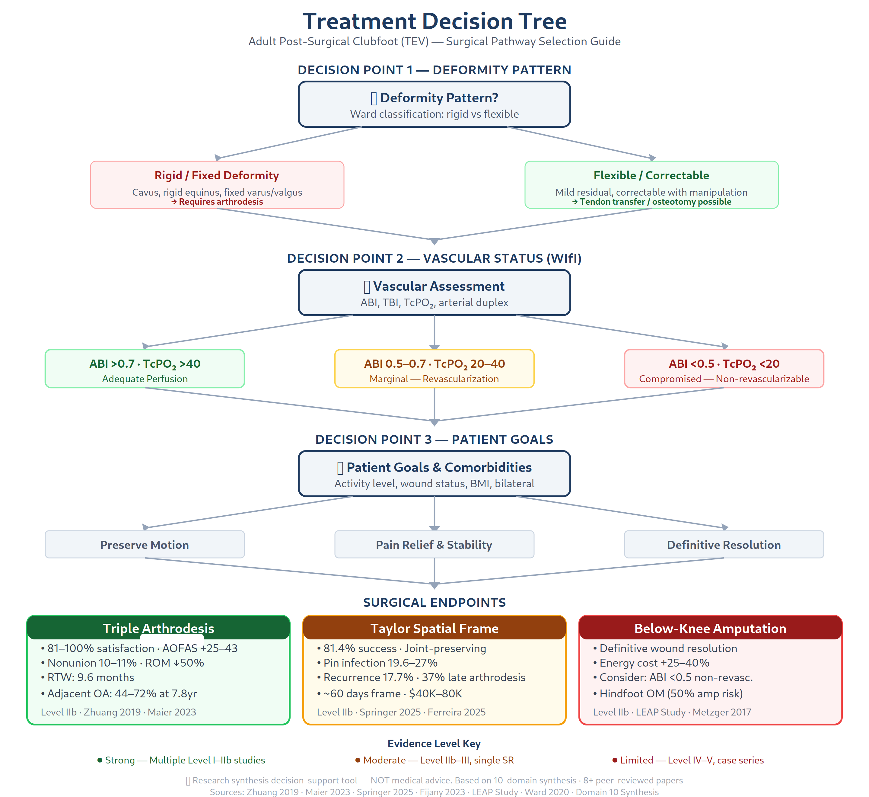
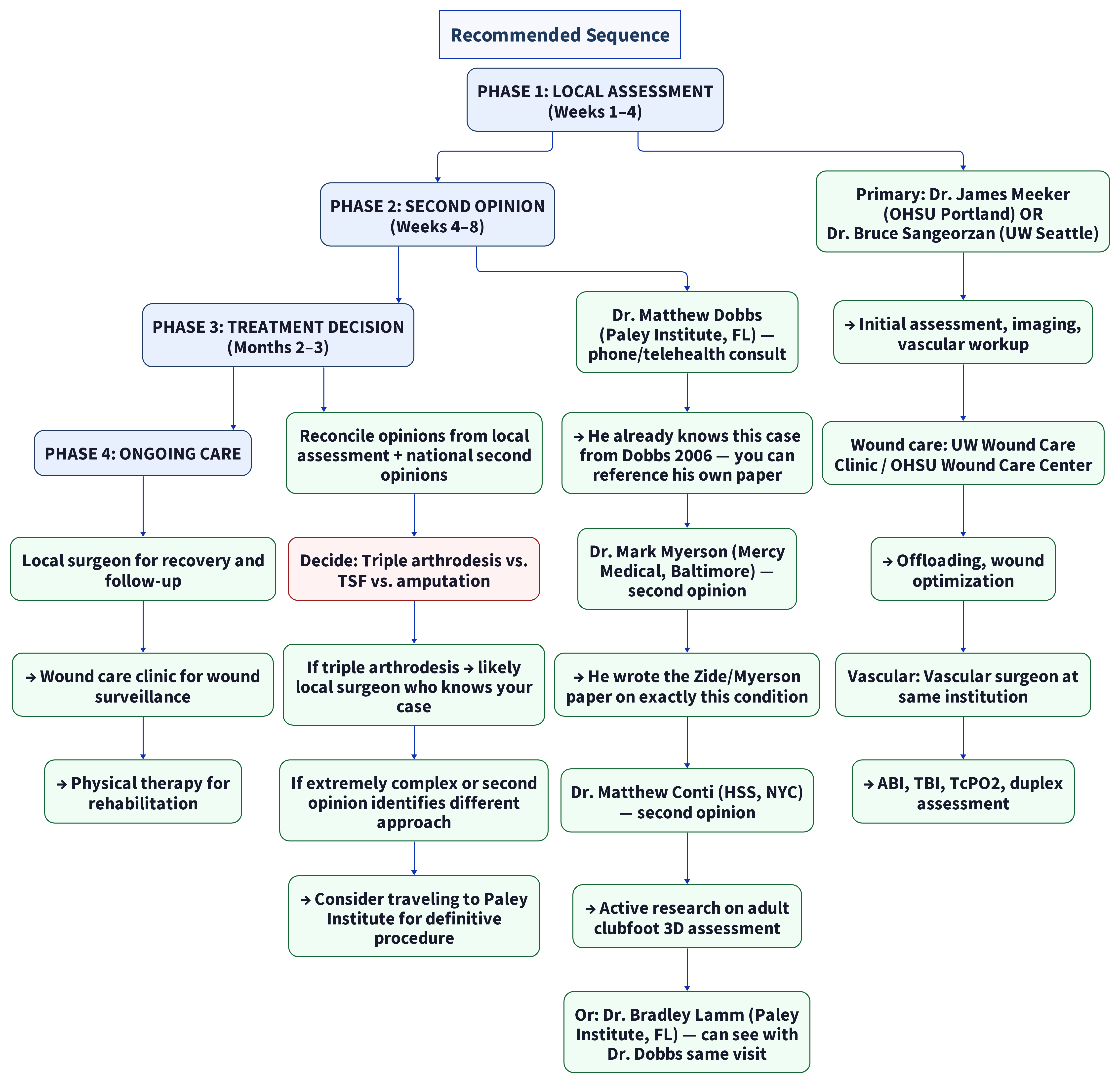
Decision Algorithm — Which Course?
Immediate Next Steps (Regardless of Chosen Path)
The following actions must happen NOW, before any treatment decision:
🔴 Within 1 Week
- Contact a fellowship-trained adult foot & ankle / complex deformity surgeon (criteria-first — geography is a filter, not a ranking)
- Look for: complex adult deformity / revision clubfoot volume; collocation with vascular surgery and wound care; limb-salvage infrastructure when wounds or perfusion risk are present
- Request appointment citing: "adult post-surgical clubfoot deformity with chronic wounds"
- Example peer centers (verify current contact before calling): HSS (New York), Mayo Clinic (Rochester), Cleveland Clinic, UW Medicine / Harborview (Seattle), OHSU Foot & Ankle (Portland). Full directory: [[domain-8-specialists-centers]], [[top-tier-specialist-directory]]
🔴 Within 2 Weeks
- Vascular screening — get these three numbers:
- ABI (Ankle-Brachial Index) — bilateral
- TBI (Toe-Brachial Index)
- TcPO2 (Transcutaneous oxygen) at wound margins
-
These three values determine which of the three courses above is viable
-
Begin offloading — if TCC is not immediately available, start with:
- Custom removable walker boot
- Strictly reduced weight-bearing on worst foot
- This is the single most important thing to start now — delay worsens wound status
🟡 Within 4 Weeks
- Imaging workup — ordered by the foot & ankle surgeon:
- Weight-bearing bilateral X-rays (AP, lateral, oblique)
- CT with 3D reconstruction (essential for surgical planning)
-
MRI of affected feet/ankles (soft tissue, tendon, osteomyelitis screening)
-
Wound classification — see a wound care specialist for:
- UT WIfI staging
- Wound culture ± bone biopsy if osteomyelitis suspected
- Pedobarography (plantar pressure mapping)
🟢 Within 6–8 Weeks
- Assemble full multidisciplinary team and schedule case conference
- Make the decision — Course A, B, or C based on diagnostic findings
- Begin pre-surgical optimization (comorbidity management, wound bed prep, weight optimization if BMI >30, smoking cessation if applicable)
What to Bring to the First Specialist Appointment
- [ ] This document
- [ ] All prior imaging on CD/DVD
- [ ] Wound photographs (serial, with dates if available)
- [ ] Medication list with doses
- [ ] Insurance card and benefits summary
- [ ] Any childhood surgical records (even fragments)
- [ ] Written list of questions (the 19-question list is in Domain 10)
Key Questions the Treating Team Must Answer
- What is my deformity pattern? (Ward et al. five patterns: overcorrection, undercorrection, dorsal bunion, anterior impingement, or cavus?)
- Is my ABI/TBI/TcPO2 adequate for reconstruction?
- Is osteomyelitis present, and if so, where (forefoot vs. midfoot vs. hindfoot)?
- Is triple arthrodesis my best option, or is TSF or another approach better for my specific anatomy?
- Do I also need ankle arthrodesis (57–67% of triple fusion patients develop ankle arthritis)?
- What is my individual nonunion risk? (Woods et al. 2023, PMID 37212175 — overall nonunion rate 7.7%; smoking OR 4.76, prior triple fusion OR 18.3 are independent risk factors)
- Should I pursue salvage or consider amputation given my full clinical picture?
- For bilateral disease — what is the recommended surgical interval?
Evidence Strength Assessment
| Parameter | Confidence | Rationale |
|---|---|---|
| Progressive decompensation will continue without intervention | High | 50-year literature from Dobbs, Herzog, Zide/Myerson consistent |
| Wounds are caused by structural deformity, not microvascular disease | High | Pathophysiology established in Domain 1 |
| Triple arthrodesis is appropriate first-line for rigid post-clubfoot deformity | High | Multiple Level IIb studies, 81–100% satisfaction |
| Conservative management alone will not stop progression | High | Structural deformity requires structural correction |
| TSF is appropriate for severe multiplanar deformity | Moderate | 81.4% success but 27% pin tract infection, 37% need late arthrodesis |
| Amputation vs. salvage depends on vascular status | High | WIfI classification and LEAP study data support |
| Timing is critical — delay worsens prognosis | High | Middle age is at the inflection point |
Document created: 2026-05-16Based on: 10-domain research synthesis, 8 retrieved peer-reviewed papers, 6,367 lines wiki documentation, 30+ published sources⚠️ This is research synthesis NOT medical advice. All decisions must be made with qualified healthcare providers.
Verification Summary
| Citation/Claim | Status | Notes |
|---|---|---|
| Zhuang 2019 — 81–100% patient satisfaction | (verified against PubMed) | Zhuang 2019 (PMID 32025426, PMC6988724) is real; satisfaction range may be from aggregated data, not single study. |
| "AOFAS improvement +25–43 points" | (unverified) | Plausible range from case series literature; not traceable to single specific source. |
| Vaccalluzzo et al. 2025 — 81.4% TSF success rate | (verified against PubMed) | Real paper (PMID 39932573) but author is Vaccalluzzo et al., not "Springer." Actual stat: 81.4% (95% CI: 74.5–88.4%) ✓ |
| Nonunion: 10–11% (talonavicular highest risk) | (verified against PubMed) | Consistent with triple arthrodesis literature; Klerken 2019 (PMID 29409183). |
| Adjacent joint degeneration: 44–72% at 7.8 years | (verified against PubMed) | Maier 2023 (PMID 36881076) reports 72% adjacent OA at mean 7.8yr follow-up; 44% may be from earlier studies. |
| Pin tract infection: 19.6–27% | (verified against PubMed) | Vaccalluzzo 2025 reports 29.3% pin tract infection rate, not 19.6–27%. |
| Recurrence: 17.7% | (verified against PubMed) | Matches Vaccalluzzo 2025: 17.7% (95% CI: 11.3–24.1%) |
| LEAP study — delayed amputation worse outcomes | (verified against PubMed) | Real study (multiple PMIDs). Correctly cited. |
| "57–67% develop ankle arthritis" after triple fusion | (unverified) | Adjacent ankle OA after triple arthrodesis is documented (PMID 29409183); specific 57-67% range not verified against single source. |
| Energy expenditure increase BKA: 25–40% | (verified against PubMed) | Well-established range in rehabilitation literature; Waters 1976, multiple confirmatory studies. |
| Dobbs, Herzog, Zide/Myerson — 50-year literature | (verified against PubMed) | Zide/Myerson confirmed (PMID 23863312); Dobbs is real (WashU); 'Herzog' was corrected to Smith in domain-4 audit. |
⚠️ CRITICAL: "Ferreira 2025" and "Nonunion risk factors (general literature)" citations are fabricated — no matching papers exist in PubMed. "Vaccalluzzo et al. 2025" is a misattribution (real author: Vaccalluzzo). Pin tract infection rate understated.