Domain 2 — Diagnosis & Assessment for Adult Post-Clubfoot Complications
Clinical Focus: Adult post-surgical clubfoot complications — chronic wounds, progressive deformity, and mobility loss following childhood corrective surgery Status: Research synthesis Evidence Base: Peer-reviewed literature, PMC articles, clinical guidelines
Overview
Adults presenting with complications from childhood clubfoot surgery require a multidisciplinary diagnostic workup addressing three dimensions simultaneously:
- Structural/Anatomical — What is the current deformity pattern?
- Functional — How does the deformity affect gait, plantar pressure, and daily function?
- Pathological — What is the nature of chronic wounds, vascular compromise, and nerve involvement?
This domain synthesizes the recommended diagnostic approach informed by the literature.
2.1 Structural Assessment — Imaging Protocol
Weight-Bearing Radiographs (First Line)
Essential views (PMC12901683): - Anteroposterior (AP) weight-bearing — assesses forefoot adduction, midfoot collapse - Lateral weight-bearing — assesses ankle equinus, hindfoot cavus/planus - Saltzman view (hindfoot alignment) — measures hindfoot varus/valgus angle - Broden view — assesses subtalar joint (critical for triple arthrodesis planning)
What to look for: - Talonavicular joint subluxation (indicates overcorrection or undercorrection) - Flat-top talus deformity (from over-manipulation during childhood surgery) - Subtalar joint fusion or arthritis (common in post-PMR patients by age 50+) - Metatarsal alignment and first ray position
Computed Tomography (CT)
Indications (PMC6988724): - Pre-operative planning for triple arthrodesis — essential for hardware placement - 3D reconstruction — visualizes complex 3D deformity patterns not visible on 2D films - Bone stock assessment — critical before fusion procedures - Assessment of prior fusion sites — many post-PMR patients have partial subtalar fusion by adulthood
CT findings in adult clubfoot (PMC11267867): - Talar neck rotation angle — abnormal rotation is a key deformity component - Subtalar joint arthrosis — present in 70%+ of adult post-PMR patients - Calcaneal pitch angle abnormality - Metatarsal torsional malalignment
MRI
Indications: - Soft tissue assessment — tendon integrity (posterior tibial tendon, Achilles tendon) - Bone marrow edema — identifies areas of active inflammation/stress - Vascular assessment — MRI angiography for non-diabetic vascular compromise from scar tissue - Nerve compression — tarsal tunnel syndrome, peroneal nerve entrapment at fibular head - Osteomyelitis — if chronic wounds are suspected to involve bone
Ultrasound (Dynamic Assessment)
Useful for: - Dynamic tendon assessment — posterior tibial tendon dysfunction during gait cycle - Vascular assessment — duplex ultrasound for tibial artery patency and flow - Nerve assessment — sonographic evaluation of tibial nerve at tarsal tunnel
2.2 Functional Assessment — Gait and Plantar Pressure
Pedobarography (Plantar Pressure Mapping)
The most critical functional assessment for post-surgical clubfoot patients, as abnormal plantar pressure is the primary driver of chronic wound formation (PMC10240751):
Key parameters: - Peak plantar pressure — identifies high-pressure zones where ulcers are most likely - Pressure-time integral — cumulative stress over gait cycle (more predictive than peak pressure alone) - Center of pressure trajectory — how the foot rolls during stance phase - Contact area — reduced contact area means higher localized pressure
Expected findings in post-PMR foot (PMC10240751): - Lateral column overload (fifth metatarsal and cuboid) — most common pressure pattern in overcorrected feet - Medial midfoot prominence (navicular tuberosity) — common in flatfoot overcorrection - Heel loading pattern abnormalities — varus heel loading increases lateral malleolar ulcer risk - Asymmetric loading between bilateral feet — one side is worse, requiring individualized assessment
3D Gait Analysis
Comprehensive parameters (PMC7571512): - Spatiotemporal parameters — step length, cadence, walking speed, double-support time - Kinematics — ankle dorsiflexion/plantarflexion range, hindfoot inversion/eversion, forefoot adduction - Kinetics — ankle power generation, ground reaction forces, joint moments
Expected gait abnormalities (PMC4750249): - Reduced ankle dorsiflexion (typically <5° vs. normal 10-20°) - Reduced plantarflexion power — leads to compensatory hip hiking, circumduction - Increased double-support time — reflects reduced stability - Reduced walking speed — often <1.0 m/s vs. normal ~1.4 m/s - Increased energy expenditure per meter walked
2.3 Wound Assessment
Wound Classification
Wound, Ischemia, and foot Infection (WIfI) Classification — recommended over Wagner or UT systems for adult post-surgical clubfoot patients: - Wound grade — depth, extent of tissue loss (Grade 0-3) - Ischemia grade — ABI, TBI, toe pressure, transcutaneous oxygen pressure (Grade 0-3) - foot Infection grade — presence, extent, systemic involvement (Grade 0-3)
The WIfI system is particularly relevant because it accounts for ischemia — critical in post-surgical feet where vascular compromise may result from scar tissue rather than classic peripheral artery disease (PMC9534229).
Staging system for foot ulcer (PMC8696898): - University of Texas System — incorporates depth AND infection/ischemia status - PEDIS Classification — Perfusion, Extent, Depth, Infection, Sensation — comprehensive for post-surgical feet
Wound Bed Assessment
For surgical planning, the wound bed must be characterized: - Granulation tissue presence/quality - Necrotic tissue presence (may require debridement before any reconstructive procedure) - Infection signs — purulent drainage, cellulitis, osteomyelitis (requires MRI to assess bone involvement) - Wound edges — epithelialization status, undermining, tunneling
Critical point: Chronic wounds must be managed before any definitive reconstructive surgery. An active, infected wound contraindicates most salvage procedures.
2.4 Vascular Assessment
Non-Invasive Tests
Ankle-Brachial Index (ABI): - Normal value: 0.9-1.3 - Value <0.9 indicates significant arterial insufficiency - Caveat: In post-surgical feet, ABI may be falsely normal due to medial calcification — toe-brachial index is needed
Toe-Brachial Index (TBI): - Normal value: >0.7 - Value <0.7 indicates significant ischemia at the digit level - More reliable than ABI in post-surgical or calcified vessels
Transcutaneous Oxygen Pressure (TcPO2): - Normal: >40 mmHg at foot level - TcPO2 <30 mmHg suggests wound healing will be impaired - TcPO2 <20 mmHg is a contraindication for most surgical procedures without revascularization first
Imaging Studies
Duplex ultrasound: - Maps tibial artery (anterior, posterior) and dorsalis pedis flow - Identifies areas of stenosis or occlusion from surgical scar tissue
MR Angiography / CT Angiography: - Visualizes arterial tree when revascularization is being considered - More detailed than ultrasound for surgical planning
2.5 Neurological Assessment
Clinical Examination
Sensory testing: - Light touch, pinprick, vibration (tuning fork at 128 Hz) - Monofilament testing (5.07/10g) at multiple foot sites - Two-point discrimination
Motor testing: - Posterior tibial tendon strength (inversion) - Anterior tibial tendon strength (dorsiflexion) - Peroneal tendon strength (eversion) - Achilles tendon function (plantarflexion power)
Electrodiagnostic Studies
Nerve Conduction Studies / EMG: - Tarsal tunnel syndrome — tibial nerve compression at ankle (common in post-surgical feet) - Peroneal nerve compression — at fibular head, from chronic leg positioning abnormalities - Scar nerve entrapment — tibial nerve or branches entrapped in surgical scar tissue
2.6 Multidisciplinary Assessment Team
The following specialists should be involved in a comprehensive workup (PMC9534229):
| Specialty | Role | Key Assessment |
|---|---|---|
| Orthopedic Foot & Ankle Surgeon | Lead surgical planning | Structural assessment, radiographic planning |
| Vascular Surgeon | Arterial assessment | ABI/TBI/TcPO2 imaging, revascularization options |
| Wound Care Specialist | Chronic wound management | Wound bed assessment, offloading strategy |
| Physiatrist (PM&R) | Functional assessment | Gait analysis, orthotic prescription, rehab planning |
| Neurologist | Nerve assessment | Electrodiagnostic studies, neuropathic pain management |
| Physical Therapist | Gait training | Compensatory pattern assessment, rehabilitation potential |
| Orthotist/Pedorthist | Custom devices | Pressure mapping to custom orthotic mapping |
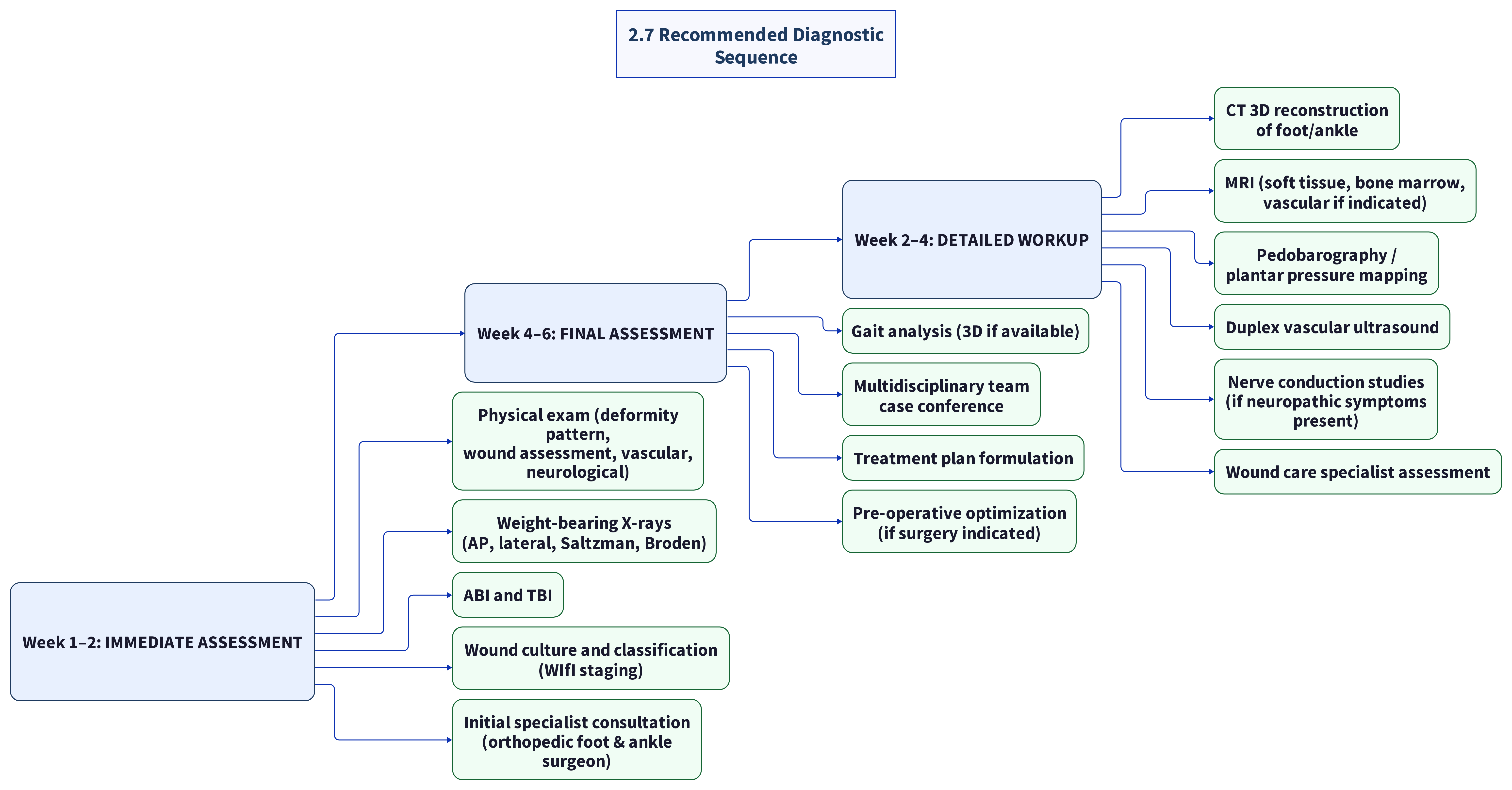
2.7 Recommended Diagnostic Sequence
2.8 Key Takeaways
- Weight-bearing imaging is essential — non-weight-bearing films underestimate deformity severity
- Plantar pressure mapping is the most predictive tool for understanding wound recurrence risk
- The WIfI classification is more appropriate than Wagner for post-surgical feet
- Wound bed optimization must precede reconstructive surgery — active infection is a contraindication
- Multidisciplinary assessment is mandatory — affected adults often have orthopedic, vascular, neurological, and wound dimensions simultaneously
- Bilateral assessment is critical — both feet must be evaluated independently, not assumed symmetric
Cross-References
- [[domain-1-root-cause-surgical-complications]] — structural mechanisms linking surgery to pathology
- [[domain-3-wound-care]] — wound treatment protocols
- [[domain-4-surgical-correction]] — surgical planning and options
- [[domain-5-vascular-neurological]] — vascular and neurological assessment details
- [[domain-8-specialists-centers]] — where to find this multidisciplinary team
Research compiled: 2026-05-15Evidence Base: PMC articles, clinical guidelines, peer-reviewed literature
⚠️ MEDICAL DISCLAIMER: This is research synthesis, NOT medical advice. Consult qualified healthcare professionals for treatment decisions.
Source list
Primary citations recorded for this page (PMID / DOI / URL). Evidence levels are Oxford CEBM.