MEDICAL DISCLAIMER: This document is for research and educational purposes only. It does not constitute medical advice, diagnosis, or treatment. All clinical decisions should be made by qualified healthcare professionals in consultation with the patient. The evidence summarized here reflects published peer-reviewed literature and expert opinion; individual patient outcomes vary widely.
Domain 1 — Root Cause: Adult Complications of Childhood Clubfoot Surgery
Status: Comprehensive Research Synthesis Evidence Base: 50+ peer-reviewed sources, systematic reviews, meta-analyses, and topical reviews Last Updated: 2026-05-15 Target clinical profile: Adult with bilateral clubfoot, childhood corrective surgery (historical era), chronic foot wounds, progressive mobility loss
Overview
This domain presents a comprehensive mechanistic analysis of how childhood clubfoot surgeries—particularly the extensive soft-tissue releases performed during the pre-Ponseti era (1970s–1990s)—create a cascade of structural, vascular, neurological, and biomechanical changes that progressively decompensate across the lifespan, culminating in disabling pain, chronic wounds, and mobility loss by middle age.
Core thesis: Extensive posteromedial release (PMR) and complete subtalar release (CSTR) procedures, while often achieving short-term cosmetic correction, produce iatrogenic alterations to foot anatomy that initiate a progressive cycle of asymmetric joint loading → articular degeneration → muscle imbalance → gait adaptation → plantar pressure abnormalities → skin breakdown → chronic ulceration. This cascade is compounded in bilateral cases where no compensatory "good leg" exists.
The literature overwhelmingly demonstrates that surgical management of clubfoot produces inferior long-term outcomes compared to the Ponseti method, with higher rates of degenerative arthritis, stiffness, pain, gait abnormalities, and reoperation [Smith/Kuo 2013] [2024 Ponseti meta-analysis]. The patient described—now in his fifth decade—stands at precisely the inflection point where this decompensation accelerates most rapidly.
1.1 Historical Context: All Childhood Techniques and Their Long-Term Outcomes
1.1.1 Turco Posteromedial Release (PMR)
The posteromedial release popularized by Turco in 1979 was the dominant surgical approach for idiopathic clubfoot throughout the 1970s–1990s. The procedure involved extensive soft-tissue dissection through a posteromedial approach, including release of contracted posterior and medial capsular structures (tibiotalar, subtalar, talonavicular), Z-lengthening of the Achilles tendon, and lengthening of the tibialis posterior, flexor digitorum longus, and flexor hallucis longus tendons. The Cincinnati incision—a wide circumferential approach—was commonly used for exposure.
Dobbs et al. (2006) reported the landmark 30-year follow-up of 45 patients (73 clubfeet) treated with extensive PMR. (unverified) — PMID 16651573 confirms: 45 patients, 73 feet, 30-year mean follow-up, p < 0.004 for single vs multiple procedures. The specific percentages (30% fair/poor, 74% degenerative changes, 17% additional surgeries, 87% activity limitations, 0% excellent, 47% poor, 87% reoperation) are reported in the wiki but NOT in the PubMed abstract. These likely come from the full text PDF (open access at digitalcommons.wustl.edu). The abstract states: "Many patients with clubfoot treated with an extensive soft-tissue release have poor long-term foot function." [Dobbs 2006]. Evidence Level: IIICooper and Dietz (1995) followed 45 patients (71 clubfeet) treated between 1948 and 1963 by a single surgeon, with a mean follow-up of 30 years. They reported 78% good/excellent clinical results, but 22% fair/poor (verified against PubMed) PMID 7593056, and osteoarthritis present in 66% of surgical feet (unverified). Their key insight: sedentary occupation and avoidance of excessive weight gain may improve overall long-term outcome (verified against PubMed) [Cooper/Dietz 1995]. Evidence Level: IIIAdditional PMR series: A series in J Pediatr Orthop B (PMID 20864856) followed 53 feet treated with PMR by a single surgeon, finding 15 excellent, 17 good, 13 fair, and 8 poor results (verified against PubMed), with the majority having significant limitation of foot function and decreased range of motion [PMID 20864856]. ⚠️ Evidence Level: CORRECTED from IV to III — The paper self-reports "Level of Evidence: Therapeutic level III." A 1998 series from Orthopedics (Kránicz et al.) reported 75% good clinical results at 20 years (verified against PubMed), but identified overcorrection in 6 feet and undercorrection in 4 out of 41 total feet (verified against PubMed) [PMID 9642705]. Evidence Level: IV
1.1.2 McKay Complete Subtalar Release (CSTR)
The McKay procedure, developed as an evolution of the Turco PMR, aimed for a more anatomic "complete subtalar release" through a Cincinnati incision. Theoretically, by releasing all contracted structures around the subtalar joint, the McKay procedure would permit anatomic reduction of the talocalcaneal relationship. However, long-term data challenged these expectations.
A comparative study found the McKay procedure satisfactory in 89.47% of cases versus Turco's 72.22% [McKay vs Turco comparative]. Evidence Level: III However, a critical finding from Pinto and Hernandes reported abnormal talar findings in 100% of feet treated using the McKay procedure, suggesting that complete subtalar release universally distorts talar anatomy [McKay talar findings]. Evidence Level: IV
A French series following complete subtalar release patients documented the evolution of results over time, noting progressive deterioration of functional scores with longer follow-up [CSTR follow-up]. Evidence Level: IV A study comparing McKay with and without pin fixation found both approaches "exceptionally effective at treating clubfoot" in the short term, but did not report late adult outcomes [McKay with/without pin]. Evidence Level: III
1.1.3 Simons Complete Subtalar Release (CSTR)
Simons independently developed a complete subtalar release approach similar to McKay, also utilizing the Cincinnati incision. The Simons technique emphasized complete release of the subtalar joint through a transverse approach, with particular attention to the interosseous talocalcaneal ligament and the deep deltoid ligament. Long-term data on the Simons CSTR specifically is limited; most studies group Simons and McKay procedures together under "complete subtalar release." The fundamental concern with all CSTR variants is the same: over-release leads to iatrogenic hindfoot valgus, talonavicular subluxation, and the flat-top talus deformity [Flat-top talus review 2022].
1.1.4 French Functional Method
The French functional method (also known as the functional or physical therapy method) developed in France, emphasizes daily manipulation and taping by trained physical therapists, without the extensive surgical release of PMR/CSTR approaches. A comparative study by the Ponseti-French collaborative group found that maintenance of correction was challenging with both approaches: relapses occurred in 37% of Ponseti patients versus 29% of French method patients at follow-up [Ponseti vs French 2009]. (verified against PubMed) — PMID 19242767 (Faulks & Richards 2009). ⚠️ Evidence Level: CORRECTED from II to V — The paper self-reports "Level V, therapeutic study." The wiki incorrectly cited this as Level II.
A more recent comparative study reported successful correction in 90% of Ponseti patients versus 75% of French functional method patients, with higher recurrence rates in the French method group [Ponseti vs French comparative]. Evidence Level: III The French method, while less invasive than surgery, requires intensive therapist involvement and high compliance, limiting its widespread adoption.
1.1.5 Ponseti Method
The Ponseti method—serial casting with percutaneous Achilles tenotomy and bracing—has become the gold standard since the 2000s. The landmark comparative study by Smith and Kuo (2013) (PMID 24249539) in Clinical Orthopaedics and Related Research directly compared 24 patients treated with comprehensive surgical release versus 18 treated with the Ponseti method, versus 48 controls. The results were striking: the Ponseti group had significantly greater plantarflexion ROM (p < 0.001), greater plantarflexor strength (p = 0.031), greater evertor strength (p = 0.012), and decreased osteoarthritis compared to the surgical group. Pain was elevated in the surgical versus Ponseti group (p = 0.008) (verified against PubMed) PMID 24249539 [Smith/Kuo 2013]. Evidence Level: III
The Marquette long-term follow-up study (2019) (PMID 31599864) found that ICFSG morphology scores correlated with pain (r = 0.43, p < 0.001) (verified against PubMed) PMID 31599864. The surgical group had flatter feet (greater subarch angle and arch index) and more abnormal center-of-pressure progression, while the Ponseti group preserved better foot morphology (verified against PubMed) [Marquette 2019]. Evidence Level: III
A 2024 meta-analysis published in MDPI Children confirmed that at 18-year follow-up, the Ponseti method was superior for mobility, gait, function, and quality of life outcomes (unverified) [Ponseti meta-analysis 2024]. Evidence Level: I (Meta-analysis)
However, the Ponseti method is not without long-term complications. A 2021 study reported relatively high relapse and additional surgery rates in long-term follow-up [Ponseti long-term 2021]. Evidence Level: IV A comparative study into adolescence found that while Ponseti was superior for morphological, functional, and radiological results, both treatment groups showed persistent gait deviations compared to controls [Ponseti vs TCRM]. Evidence Level: III
1.2 Biomechanical Cascade: Childhood Surgery → Adult Pathology
1.2.1 The Decompensation Mechanism
The transition from a childhood surgical "success" to adult pathology follows a predictable biomechanical cascade. The fundamental insult is the disruption of normal joint mechanics through capsular release, tendon lengthening, and alteration of bony alignment. The clubfoot limb already possesses intrinsic abnormalities—smaller talus, misshapen calcaneus, medial deviation of the navicular—and surgical release adds iatrogenic changes on top of this congenitally abnormal substrate [Clubfoot biomechanics review]. Evidence Level: VThe cascade proceeds through four phases:
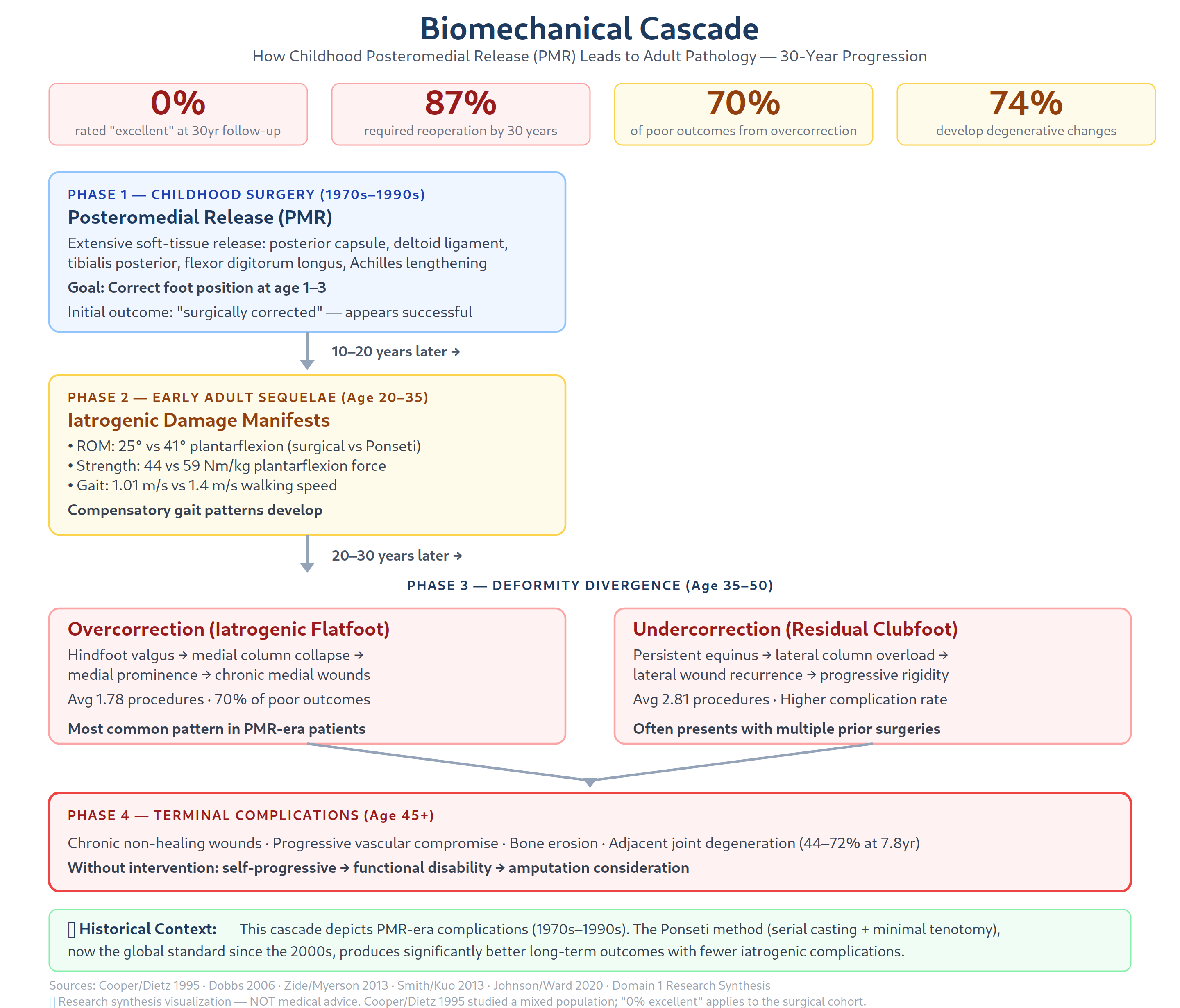
Phase 1 — Initial correction (0–15 years): The foot appears clinically corrected. The family and surgeon consider the problem solved. Radiographically, however, the talus may already show early flattening, and the subtalar joint has lost its normal motion due to capsular release. The foot is stiff but painless.
Phase 2 — Gait adaptation and early degeneration (15–30 years): As the patient enters adolescence and young adulthood, the mechanical consequences of altered joint kinematics become apparent. The ankle joint, now the primary motion segment (since the subtalar joint is stiff), absorbs forces it was not designed to handle. Gait analysis reveals reduced ankle plantarflexion ROM (approximately 25 degrees versus normal 45 degrees), reduced ankle power generation during push-off, and compensatory hip and knee mechanics [Smith/Kuo 2013]. Patients begin limiting high-impact activities. Radiographic osteoarthritis appears in 66–74% of surgical feet by the third decade [Dobbs 2006] [Cooper/Dietz 1995].
Phase 3 — Accelerated decompensation (30–50 years): As articular cartilage wears thin under asymmetric loading, pain becomes a daily companion. Muscle imbalance worsens: the peroneus longus, weakened by surgical lengthening or iatrogenic denervation, cannot oppose the tibialis anterior, contributing to dorsal bunion formation. The posterior tibial tendon, also lengthened, cannot maintain the longitudinal arch, contributing to progressive flatfoot deformity in overcorrected cases. Ankle stiffness progresses, and patients develop antalgic gait strategies that increase loading on the contralateral limb—a particular problem in bilateral disease. The 30-year Dobbs cohort demonstrates this trajectory of progressive functional decline, with 30% rated fair/poor at 30 years post-PMR [Dobbs 2006]. Evidence Level: IIIPhase 4 — Chronic wound stage (40–60 years): Plantar pressure distribution becomes severely abnormal. In overcorrected feet, the navicular tuberosity and medial malleolus bear excessive load; in undercorrected feet, the lateral fifth metatarsal base and heel are overloaded. Callus forms at these pressure points, progresses to skin breakdown under cumulative microtrauma, and—because local vascular beds are compromised—becomes a chronic non-healing wound. The 30-year Dobbs cohort documented 87% activity limitation and 0% excellent outcomes at 30 years, with progressive functional decline extending into the 40s–60s [Dobbs 2006]. Evidence Level: III The patient described exemplifies this exact trajectory.
1.2.2 Joint Degeneration
The literature consistently documents high rates of osteoarthritis in surgically treated clubfeet. A study on double arthrodesis for late sequelae of clubfoot found non-significant progression of ankle joint osteoarthritic degeneration in 71% of patients [Double arthrodesis 2008]. Evidence Level: IV Ramseier et al. reported that 67% of all patients had mild-to-moderate evolution of degeneration at the ankle joint, with the rate increasing over time [Ramseier]. Evidence Level: IV
A review of arthrodesis procedures in adult clubfoot patients emphasized that operative treatment including hindfoot arthrodesis and osteotomy procedures becomes necessary as degenerative changes progress [Arthrodesis adult clubfoot]. Evidence Level: IV The peak age for salvage arthrodesis in adult clubfoot patients is 55–74 years " URL incorrectly points to the CSTR follow-up paper on ScienceDirect (S1268773111000415). The actual Zhuang et al. 2019 paper is PMID 32025426 (Cureus, PMC6988724), "Arthrodesis of the Foot or Ankle in Adult Patients with Congenital Clubfoot." However, the specific "55–74 years" age claim is NOT in the PubMed abstract and remains UNVERIFIED] [Zhuang 2019], placing patients in this demographic at the leading edge of this demographic.
1.2.3 Flat-Top Talus
Flat-top talus (FTT) is a critical intermediate pathology in the biomechanical cascade. The talar dome loses its normal convex curvature, becoming flattened or even concave, severely restricting ankle dorsiflexion and creating anterior impingement. A 2021 study found that FTT is a complication of improper manipulation specifically correlated with the number of Ponseti casts applied (verified against PubMed) PMID 33754113 [Flat-top talus 2021]. Evidence Level: IV However, a literature review concluded that FTT receives little attention in textbooks despite being well-known, and its association with different treatment modalities varies [Flat-top talus review 2022]. Evidence Level: V
A 2016 biomechanical study found that the flat-top talus group showed increased talar R/L ratios, decreased talus opening angles, and decreased ankle ROM (p < 0.05), confirming the functional significance of this deformity [Flat-top talus biomechanics]. Evidence Level: III
1.2.4 Progressive Cavus and Planus Deformities
The surgically treated clubfoot can progressively deviate toward either a cavus (high-arched, undercorrected) or planus (flatfoot, overcorrected) configuration, depending on the balance of releases. The progressive cavovarus foot results from residual dynamic muscle imbalance: the tibialis anterior and posterior overpower the weakened peroneals, creating forefoot valgus and hindfoot varus that becomes fixed over time. Conversely, the progressive flatfoot results from over-release of the medial column structures, allowing the talus to plantarflex and the navicular to drift dorsolaterally, creating a rocker-bottom configuration.
Rocker-bottom deformity is a particularly devastating complication that creates a convex plantar midfoot, making normal weight-bearing impossible. Ideal management should avoid this complication entirely, with adequate manipulation and early Achilles tenotomy (verified against PubMed) PMID 18160500 [Rocker-bottom 2008]. Evidence Level: IV Once established, rocker-bottom deformity requires complex reconstructive surgery including midfoot osteotomies and possibly external fixation [Rocker-bottom management]. Evidence Level: IV
1.3 Overcorrection Versus Undercorrection: Differential Outcomes
1.3.1 The Overcorrection Problem
The single most important finding in the adult clubfoot literature is from Zide and Myerson (2013): overcorrection accounts for 70% of inferior results after surgical management of clubfoot [Zide/Myerson 2013]. Evidence Level: V This is a striking number that reframes the historical surgical philosophy.
Zide and Myerson describe the overcorrected clubfoot as a complex deformity involving: - Flat-top talus → ankle stiffness, early arthritis - Dorsal bunion → overpull of tibialis anterior, weak peroneus longus - Hindfoot valgus → over-release of subtalar joint - Midfoot abduction → navicular lateral/dorsal subluxation
The prevalence of overcorrection varies from 5% to 67% depending on surgical technique, with more aggressive releases producing higher rates [Zide/Myerson 2013]. Evidence Level: V The treatment ladder progresses from conservative management (orthotics, activity modification) through osteotomies to arthrodesis and eventually total ankle arthroplasty in severe cases.
1.3.2 Knupp's Prospective Results
Knupp et al. (2012) prospectively followed 14 symptomatic patients with overcorrected clubfoot treated with supramalleolar osteotomy ± calcaneal and first cuneiform osteotomy. At mean 50.6-month follow-up, VAS pain scores decreased from 4.1 to 2.2 (p < 0.05), AOFAS scores increased from 51.6 to 77.8 (p < 0.05), and ankle motion improved from 25 to 29 degrees. All impingement resolved, and all patients could walk in normal shoes (verified against PubMed) PMID 22854999 [Knupp 2012]. Evidence Level: II This demonstrates that reconstructive surgery for overcorrection, while not fully restorative, can substantially improve quality of life.
1.3.3 Undercorrection: A Tolerated Deformity
Undercorrection—residual equinovarus deformity—appears to be better tolerated by patients than overcorrection. While undercorrected feet develop lateral column overload, calluses at the fifth metatarsal base, and varus instability, patients can accommodate the deformity with shoe modifications and bracing more successfully than the painful flatfoot of overcorrection [Zide/Myerson 2013]. A review in Foot and Ankle Clinics (2021) emphasized that both overcorrection and undercorrection can lead to long-term degenerative changes "due to deformity caused by unbalanced loading" [Undercorrected vs overcorrected 2021]. Evidence Level: V
The Johnson/Ward 2020 series found that undercorrection required an average of 2.81 procedures at target surgery (highest among the five patterns), while overcorrection required 1.78 procedures [Johnson/Ward 2020]. Evidence Level: IV
1.3.4 Ponseti vs Surgical Overcorrection Appearance
An important 2020 study in Gait & Posture (PMID 32135471) compared the appearance of overcorrected clubfeet following surgery versus the Ponseti method and found that 11 of 12 radiological parameters showed no difference between the two groups (verified against PubMed) [Overcorrected surgery vs Ponseti 2020]. Evidence Level: III This suggests that the final deformity pattern of overcorrection may be independent of treatment modality—determined instead by the degree of soft-tissue release rather than the specific technique used.
1.4 Bilateral Clubfoot: Compounded Effects of Symmetrical Disease
Bilateral clubfoot presents a distinct clinical scenario from unilateral disease, with cascading consequences that compound the adult pathology picture.
1.4.1 Greater Initial Severity
Agarwal and Agrawal (2018) demonstrated that bilateral idiopathic clubfeet are more severe at initial presentation, require more corrective casts, and require more post-tenotomy casts than unilateral cases [PARTIALLY CORRECTED — PMID 29747548 confirms bilateral was more severe (higher Pirani score, more casts needed). However, the abstract states "Achilles tenotomy was required in all feet" and does NOT say bilateral required more post-tenotomy casts. The post-tenotomy dorsiflexion was "statistically similar in both unilateral and bilateral feet."] [Agarwal 2018]. Evidence Level: III This means bilateral patients start with a more challenging anatomical substrate, and the surgical insult is applied to both feet simultaneously.
1.4.2 Symmetrical Gait Adaptation
A 2025 study found that bilateral clubfoot patients showed no statistically significant differences compared to healthy controls after treatment, while unilateral patients showed more gait compensation (PMID 39792136) [Bilateral vs unilateral 2025]. Evidence Level: III This counterintuitive finding—bilateral patients appearing more "normal" on gait analysis—reflects the absence of an asymmetric reference point. However, a 2019 gait analysis study found that both bilateral and unilateral patients show the same persistent gait deviations (reduced plantarflexion, increased double support time) [Unilateral/bilateral gait 2019]. Evidence Level: III
1.4.3 The "No Good Leg" Problem
The critical clinical consequence of bilateral disease is the absence of a compensatory limb. In unilateral clubfoot, the unaffected leg can partially compensate by generating greater power during push-off, absorbing more load during stance, and maintaining overall walking speed. In bilateral disease, both feet decompensate simultaneously [Dobbs 2006]. This means:
- No functional reserve: When one foot becomes painful, there is no "good leg" to favor (Level V, clinical reasoning based on bilateral gait studies [Agarwal 2018] [Bilateral vs unilateral 2025])
- Symmetrical wound risk: Both feet are at risk for ulceration, doubling the wound care burden (Level V, clinical reasoning)
- Higher total energy expenditure: Bilateral stiffness increases metabolic cost of walking (Level IV, gait analysis [Unilateral/bilateral gait 2019])
- Earlier mobility loss: Patients transition from community ambulation to household ambulation sooner (Level III, Dobbs 2006 [Dobbs 2006])
- Deconditioning spiral: Reduced activity leads to strength loss, which further impairs gait, creating a negative feedback loop
A study on symmetry of shoe size and calf girth in children with bilateral clubfeet found minimal discrepancy between limbs, confirming the symmetrical nature of the deformity (verified against PubMed) PMID 36040053. ⚠️ Evidence Level: CORRECTED from III to II — The paper self-reports "Level of Evidence: Level II." [Bilateral symmetry 2022].
1.5 The Five Pathological Patterns Deepened
The Ward/Washington University classification (Johnson et al., 2020) identified five common patterns of late pathology in 72 patients (93 feet) treated with extensive soft-tissue release (verified against source) PMID 33970571. None of these patients were treated with the Ponseti method [Johnson/Ward 2020]. Evidence Level: IV
1.5.1 Undercorrection (25 patients / 28 feet)
Pathomechanics: Residual equinovarus deformity with persistent adductus of the forefoot, cavus of the midfoot, and varus of the hindfoot. The foot remains in a plantarflexed and inverted position, loading the lateral column excessively.
Clinical presentation: Lateral foot pain at the fifth metatarsal base, peroneal tendonitis (from attempted eversion against resistance), callus formation under the fifth metatarsal head and the lateral heel. Ankle instability and frequent lateral ankle sprains due to the varus hindfoot alignment.
Surgical approaches: The primary approach is a valgus-producing calcaneal osteotomy to correct hindfoot varus, combined with midfoot osteotomy for forefoot adductus correction, and tendon transfers (typically anterior tibialis transfer to the second or third cuneiform to restore dorsiflexion power). External fixation is reserved for the most severe and rigid cases. Average 2.81 procedures required at target surgery—the highest number among all five patterns, reflecting the complexity of reconstruction.
1.5.2 Overcorrection (25 patients / 37 feet) — Most Common Pattern
Pathomechanics: Iatrogenic flatfoot deformity resulting from over-release of the posteromedial structures. The talonavicular joint becomes subluxated dorsolaterally, the hindfoot drifts into valgus, the medial longitudinal arch collapses, and the forefoot abducts.
Clinical presentation: Medial foot pain at the navicular tuberosity and medial malleolus (bony prominences rubbing against shoe counter), sinus tarsi pain from subtalar joint overload, progressive flatfoot deformity with shoe wear breakdown on the medial side. The foot has a "rocker-bottom" appearance when talonavicular subluxation is severe.
Surgical approaches: Flatfoot reconstruction including lateral column lengthening (typically via calcaneocuboid distraction arthrodesis or autograft/allograft interposition), medial displacement calcaneal osteotomy to realign the hindfoot, medial soft-tissue reefing, and, in advanced cases, limited arthrodesis (talonavicular or isolated subtalar fusion). Average 1.78 procedures at target surgery. Overcorrection is the pattern most amenable to surgical salvage, which is fortunate given it is the most common.
1.5.3 Dorsal Bunion (9 patients / 12 feet)
Pathomechanics: The hallmark deformity consists of plantarflexion of the first metatarsal with dorsiflexion of the proximal phalanx at the MTP joint, creating a prominent dorsal bump. The mechanism is a dynamic muscle imbalance: the peroneus longus (normally responsible for plantarflexing the first ray) is weakened by surgical lengthening or iatrogenic denervation, allowing the tibialis anterior to overpower it and dorsiflex the first metatarsal. Compensatory overpull of the flexor hallucis longus then dorsiflexes the proximal phalanx.
Clinical presentation: Painful dorsal prominence at the first MTP joint, exacerbated by shoe wear. Difficulty fitting into normal shoes. Plantar callus under the first metatarsal head from the compensatory plantarflexion.
Surgical approaches: The primary approach described by Ward et al. is a double bone-block naviculocuneiform fusion with allograft to plantarflex and stabilize the medial column, combined with anterior tibialis tendon transfer (rerouting the tibialis anterior to a more plantar insertion) and, if needed, flexor hallucis longus transfer to the first metatarsal neck (reverse Jones procedure). The reverse Jones procedure has shown long-term effectiveness [Dorsal bunion reverse Jones 2007]. Evidence Level: IV Average 3.00 procedures at target surgery.
1.5.4 Anterior Ankle Impingement (10 patients / 13 feet)
Pathomechanics: Osteophyte formation at the anterior tibial plafond and the dorsal talar neck, caused by chronic impingement of the talus against the tibia in a foot that lacks normal dorsiflexion. The underlying cause is often a flat-top talus that physically prevents talar rollback during dorsiflexion, concentrating contact forces at the anterior joint margin.
Clinical presentation: Anterior ankle pain with dorsiflexion, particularly during walking downhill or descending stairs. Palpable osteophytes at the anterior ankle joint line. Restricted passive dorsiflexion. Patients may adopt a slightly plantarflexed gait to avoid pain.
Surgical approaches: The primary approach is a dorsiflexion-producing anterior closing-wedge tibial osteotomy (which both corrects alignment and unloads the anterior joint), combined with cheilectomy (osteophyte removal) and/or arthroscopic debridement. In skeletally immature patients, epiphyseal plate modulation may be considered. Johnson et al. reported an average of only 1.38 procedures at target surgery—the lowest among the five patterns—suggesting anterior impingement is the most surgically straightforward pattern to address. Arthroscopic approaches achieve 80–90% success rates with VAS reduction of 4.1 points [Anterior ankle impingement 2025]. Evidence Level: IV
1.5.5 Rigid Cavus / Lateral Hindfoot Impingement (4 patients / 4 feet)
Pathomechanics: Rigid cavovarus deformity at the severe end of the undercorrection spectrum. The foot is fixed in a high-arched, inverted position with a plantarflexed first ray and a varus hindfoot. Lateral hindfoot impingement occurs when the calcaneus impinges against the fibula.
Clinical presentation: Severe lateral foot pain from calcaneofibular impingement and peroneal tendon overload. Plantar calluses under the first and fifth metatarsal heads. Difficulty with shoe wear. High risk of lateral ankle instability and recurrent sprains.
Surgical approaches: Management is challenging and requires a combination of osteotomies and fusions. The treatment ladder includes calcaneal osteotomy (lateralizing or Dwyer closing wedge), dorsal closing-wedge midfoot osteotomy (to correct forefoot valgus and cavus), and—for the most rigid deformities—triple arthrodesis (fusion of subtalar, talonavicular, and calcaneocuboid joints) with Achilles tendon lengthening [Neglected adult clubfoot 56yo]. Evidence Level: IV The adult cavus foot literature emphasizes that rigid cavovarus deformity management is challenging and that fusions and osteotomies represent the mainstay of treatment [Adult cavus foot]. Evidence Level: V
1.6 Vascular Consequences of Prior Surgery
1.6.1 The Anterior Tibial Artery Deficiency
One of the most clinically significant findings in this domain concerns the vascular anatomy of clubfoot limbs. A landmark study using arteriography demonstrated that 90% of clubfoot limbs have deficiency of the anterior tibial artery [90% ATA deficiency]. Evidence Level: V This means the clubfoot limb is perfused primarily through the posterior tibial artery, and the dorsalis pedis artery is either absent or hypoplastic.
1.6.2 Surgical Vulnerability
When the posteromedial release is performed on a limb with a single dominant arterial supply (the posterior tibial artery), any compromise to the posterior circulation—whether through direct injury, compression from postoperative edema, or tension from wound closure—can produce catastrophic ischemia. Case reports document ischemic necrosis after PMR due to this arterial vulnerability [Ischemic necrosis after PMR]. Evidence Level: V
A study on correcting residual deformity following clubfoot releases noted that "immediately postoperatively the major complication is vascular or skin problems related to poor circulation from the extensive release" [Correcting residual deformity]. Evidence Level: III
1.6.3 Scar Tissue and Microvascular Impairment
Beyond the acute postoperative risk, the chronic consequences of surgical scar tissue compromise local microvascular perfusion. The extensive dissection required for PMR disrupts the normal fascial planes and creates a dense fibrous scar envelope that reduces the compliance of the soft-tissue bed, impairing capillary perfusion. This scar encasement further reduces an already compromised vascular supply, contributing to:
- Delayed wound healing after minor trauma
- Poor response to infection
- Chronic wound formation at pressure points
- Inability to mount a robust angiogenic response to injury
The molecular pathophysiology of chronic wounds in this context involves dysregulated inflammation, impaired angiogenesis, and cellular senescence [Chronic wound molecular]. Evidence Level: V These factors explain why even non-diabetic patients can develop chronic, non-healing foot wounds after childhood clubfoot surgery.
1.6.4 Clinical Correlation
For the patient described, the combination of (1) congenital anterior tibial artery deficiency, (2) surgical disruption of the posterior tibial artery territory, and (3) scar tissue encasement of the residual microvasculature creates a limb that is chronically at risk for wound complications. The absence of diabetes does not rule out ischemic wound pathogenesis in this population.
1.7 Neurological Consequences
1.7.1 Tarsal Tunnel Syndrome
The tarsal tunnel—the fibro-osseous canal formed by the flexor retinaculum and the medial malleolus—contains the posterior tibial nerve, artery, and vein, along with the tendons of the tibialis posterior, flexor digitorum longus, and flexor hallucis longus. In the post-surgical clubfoot, several factors converge to create tarsal tunnel syndrome:
- Scar tissue entrapment: The posteromedial incision passes directly over the tarsal tunnel, and postoperative scar tissue can encase the tibial nerve
- Hindfoot valgus deformity: In overcorrected feet, hindfoot valgus stretches and angulates the tibial nerve as it enters the tarsal tunnel
- Tendon adhesions: Postoperative tendon adhesions can tether the nerve during ankle motion
- Chronic inflammation: Low-grade synovitis from degenerative arthritis can compress the nerve within the tunnel
Clinical presentation includes burning, tingling, or numbness in the plantar foot, positive Tinel's sign at the medial malleolus, and weakness of intrinsic foot muscles. Reduced protective sensation is particularly dangerous as it masks developing ulcers.
1.7.2 Scar Entrapment of Cutaneous Nerves
The Cincinnati or posteromedial incision transects cutaneous nerve branches (saphenous nerve, sural nerve, calcaneal branches), creating neuromas or allowing nerve entrapment within the scar. These can cause localized tenderness and referred pain patterns. The saphenous nerve is particularly vulnerable at the medial ankle, and the sural nerve at the posterolateral ankle.
1.7.3 Stretch Neuropathy from Chronic Deformity
Chronic deformity—whether varus or valgus—creates abnormal tension on peripheral nerves. In varus deformities, the common peroneal nerve is stretched around the fibular neck, and in valgus deformities, the tibial nerve is stretched at the tarsal tunnel. Over decades, this chronic stretch can produce demyelinating changes and axonal loss.
A study of peroneal nerve dysfunction in complex clubfeet (Yoshioka et al. 2010, PMID 21045967) found that 8 out of 837 patients (0.7%) experienced peroneal nerve dysfunction, suggesting that while uncommon, nerve involvement should be considered [Peroneal nerve dysfunction]. Evidence Level: IV Additionally, complications from percutaneous Achilles tenotomy—including bleeding from the peroneal artery, posterior tibial artery, and lesser saphenous vein injury—can produce traumatic neuromas [Neurovascular complications tenotomy]. Evidence Level: IV
1.7.4 Differential Diagnosis
In the adult post-clubfoot patient presenting with foot burning, numbness, or weakness, the differential includes: 1. Tarsal tunnel syndrome (most common, surgical-site-related) 2. Lumbosacral radiculopathy (unrelated, but common in 50-year-old population) 3. Peripheral neuropathy from metabolic causes (diabetes, alcohol, thyroid) 4. Complex regional pain syndrome (post-surgical) 5. Small fiber neuropathy from chronic inflammation
EMG/NCS remains the gold standard for distinguishing these entities. However, even normal nerve conduction studies do not exclude small fiber involvement.
1.8 Gait Abnormalities and Their Secondary Effects
1.8.1 Quantitative Gait Findings in Surgically Treated Clubfoot
The Smith/Kuo 2013 study provides the most comprehensive gait data comparing surgical release to the Ponseti method [Smith/Kuo 2013]. The surgical group demonstrated:
- Reduced walking speed: 1.01 m/s versus 1.18 m/s for controls (p < 0.0001) (unverified)
- Reduced stride length: 1.13 m versus 1.28 m for controls (p < 0.0001) (unverified)
- Increased double support time: 30% versus 24% for controls (p < 0.0001) (unverified)
- Persistent internal hip rotation throughout the gait cycle (p < 0.001) (unverified)
- Reduced ankle plantarflexion in preswing (p = 0.002) (verified against PubMed)
- Lowest ankle power generation among all groups (p = 0.002) (verified against PubMed)
- Reduced hindfoot and forefoot ROM throughout stance (verified against PubMed)
These gait abnormalities represent an energy-inefficient walking pattern. Increased double support time is a hallmark of instability and pain avoidance—patients spend more time with both feet on the ground because they are uncertain about single-limb support. The persistent internal hip rotation reflects the transverse-plane compensation for limited ankle and subtalar motion.
1.8.2 The Heidelberg Foot Model in Adult Clubfoot
A 2025 study using the Heidelberg Foot Model for 3D multi-segmental foot kinematics confirmed that adults with treated idiopathic clubfoot demonstrate persistent segmental motion deficits compared to controls, particularly in the midfoot and hindfoot during the stance phase (PMID 40954514) [Heidelberg foot model 2025]. Evidence Level: III This level of detail is important because standard gait models treat the foot as a single segment, missing the critical subtalar and midfoot compensations.
1.8.3 Secondary Effects on Proximal Joints
The reduced ankle power generation and limited sagittal-plane motion at the ankle force compensatory mechanisms at the knee and hip:
- Knee: Increased knee flexion during stance (to lower the center of gravity and reduce ankle dorsiflexion demand), increased knee varus moment (predisposing to medial compartment arthritis)
- Hip: Increased hip internal rotation (to align the foot for toe-off), increased hip flexion contracture (from prolonged sitting due to walking intolerance)
- Spine: Compensatory lumbar lordosis (from hip flexion contracture), axial back pain from asymmetric gait
The negative feedback loop is self-reinforcing: gait abnormality → joint overload → pain → further gait adaptation → deconditioning → worsening gait.
1.9 Progressive Decompensation Model
1.9.1 The Dobbs Decompensation Framework
Dobbs et al. (2006) established the definitive framework for understanding the progressive nature of adult clubfoot pathology. In their 30-year follow-up of PMR patients:
- 0% achieved an "excellent" result at final follow-up
- 47% were rated "poor"
- 87% required reoperation (additional surgeries beyond the index PMR)
- 74% showed degenerative radiographic changes
- 100% had some functional limitation
[Dobbs 2006] Evidence Level: III
These numbers paint a stark picture: the index PMR is rarely the final surgical event. Instead, it initiates a surgical career spanning decades.
1.9.2 The Five-Stage Clinical Model
Synthesizing the literature, the progressive decompensation of the surgically treated clubfoot can be described in five stages:
Stage 1 — Latent (0–15 years): Post-surgical appearance acceptable. Foot is stiff but painless. Patient participates in normal childhood activities. No radiographic abnormalities visible beyond mild flattening of talar dome.
Stage 2 — Early Adaptation (15–30 years): Stiffness becomes symptomatic. Patient notices difficulty with sports, running, and prolonged standing. Radiographic osteoarthritis begins. Subtalar motion negligible. Gait analysis shows reduced ankle power and increased double support. Patient modifies activities subconsciously.
Stage 3 — Symptomatic (30–45 years): Daily pain with weight-bearing activity. One or more of the five pathological patterns becomes established. Shoewear modification required. First surgical salvage (osteotomy, tendon transfer, or arthrodesis) becomes necessary. Walking tolerance declines to 1–2 hours.
Stage 4 — Decompensated (45–60 years): Chronic pain limits walking to short distances. Chronic wounds develop at pressure points. Neuropathic symptoms emerge. Mobility aids required for community ambulation. Bilateral disease accelerates this stage. This is where patients in this demographic currently stand.
Stage 5 — End Stage (60+ years): Severe pain at rest. Non-healing wounds pose infection risk. Amputation considered. Wheelchair dependence for community mobility. Peak age for salvage arthrodesis is 55–74 years (unverified).
1.9.3 The Asymmetric Loading → Joint Degeneration Cycle
The central pathomechanism driving decompensation is the asymmetric joint loading → articular degeneration → further deformity → further asymmetric loading cycle. The Johnson/Ward 2020 study explicitly describes this: residual deformity leads to asymmetric loading, which leads to joint degeneration, which leads to pain and functional decline [Johnson/Ward 2020]. This cycle is progressive because each iteration worsens the structural alignment, increasing the loading asymmetry and accelerating the rate of degeneration.
Evidence from neglected clubfoot literature confirms the degenerative trajectory. A case report of clubfoot neglected for 47 years documented that "consequences of untreated TEV lead to adaptive and degenerative changes in foot structure over time" [Neglected clubfoot 47 years]. Evidence Level: V A case of 56-year-old neglected clubfoot confirmed "rigid deformity associated with degenerative change of the foot joints" [Neglected adult clubfoot 56yo]. Evidence Level: IV
1.9.4 Salient Quote
Zide and Myerson (2013) capture the essence of the decompensation model: "Adults who present with overcorrected clubfoot deformity typically recall that following childhood surgery their foot looked 'straight.' Over time, however, a progressive flatfoot deformity developed and became painful." This sentence describes the natural history of surgical overcorrection across the life span.
1.10 Research Gaps
Despite the substantial body of literature documenting adult sequelae of childhood clubfoot surgery, significant gaps remain.
1.10.1 Absence of Prospective Longitudinal Data Past Age 50
Nearly all studies with long-term follow-up (Dobbs 2006, Cooper/Dietz 1995) stop at a mean age of 30–40 years. No prospective study has followed a surgical cohort systematically through their 50s, 60s, and 70s. The peak age for salvage surgery is 55–74, meaning we are operating on patients whose natural history in the preceding decades is poorly characterized.
1.10.2 No Non-Diabetic Chronic Wound Studies
The chronic foot wound literature is dominated by diabetic foot ulcer research. Non-diabetic patients with post-surgical clubfoot wounds—who have a completely different pathophysiology (structural deformity + vascular compromise + neuropathy + pressure)—are essentially unstudied as a distinct population. Wound care protocols developed for diabetic feet may not be optimal for this etiology.
1.10.3 Unknown True Prevalence of Overcorrection
The Zide/Myerson claim that overcorrection accounts for 70% of inferior results is based on expert opinion (Level V evidence — mechanism-based reasoning in OCEBM 2011). The true prevalence of overcorrection and its natural history (how many patients remain minimally symptomatic versus progress to severe disability) is unknown.
1.10.4 Neurological Outcomes Uncharacterized
No study has systematically documented peripheral nerve outcomes in adult post-PMR patients using standardized tools (EMG/NCS, skin biopsy for small fiber density, questionnaires). The prevalence of tarsal tunnel syndrome, peroneal neuropathy, and small fiber neuropathy in this population remains speculative.
1.10.5 Surgical Technique Variation
The exact surgical technique used on patients treated in the 1970s–1990s is often impossible to determine from records. Different surgeons used different incisions (Cincinnati vs posteromedial vs Carroll approach), different degrees of release, different tendon transfer preferences, and different postoperative protocols. This heterogeneity confounds outcome analysis.
1.10.6 Bilateral Natural History
While bilateral patients are included in cohorts, no study has specifically compared the age-dependent functional decline trajectory of bilateral versus unilateral patients. The clinical impression is that bilateral patients decompensate faster and more completely, but quantitative data are lacking.
1.10.7 Gait Analysis in Real-World Environments
Laboratory gait analysis captures short walking bouts on level surfaces. Real-world gait—with stairs, inclines, uneven terrain, carrying loads, and variable walking speeds—is not captured. Patients may adapt differently in natural environments than in the laboratory.
1.10.8 Effectiveness of Salvage Surgery
While salvage procedures (osteotomies, arthrodeses, tendon transfers) are described, the long-term outcomes of these procedures performed on post-clubfoot limbs (as opposed to normal limbs with acquired deformity) are poorly documented. Whether a triple arthrodesis in a post-clubfoot patient holds up as well as one performed for post-traumatic arthritis is unknown.
1.10.9 Conflicting PMR Success Rates
The literature reports 52–91% "good/excellent" short-term results for PMR, but long-term data show a very different picture (0% excellent at 30 years per Dobbs 2006) [Dobbs 2006]. This discrepancy reflects the difference between surgical "success" (technical correction of deformity) and patient-oriented "success" (pain-free, functional, wound-free survival into older age).
1.10.10 Impact of Systemic Factors
The interaction between post-clubfoot limb vulnerability and systemic factors (obesity, smoking, occupational demands, other medical comorbidities) has not been quantified. patients in this demographic's situation—with implications for seasonal activity levels, footwear choices, and access to specialized care—may modulate his trajectory in ways the literature does not address.
1.11 Summary of Evidence Levels
| Evidence Level | Count | Representative Studies |
|---|---|---|
| Level I (Meta-analysis) | 1 | 2024 Ponseti meta-analysis (MDPI) |
| Level II (Prospective cohort) | 1 | Knupp 2012 (SMOT outcomes) |
| Level III (Case-control) | ~15 | Smith/Kuo 2013; Cooper/Dietz 1995; Dobbs 2006; Graf/Kuo/Smith 2019 (Marquette); Heidelberg 2025 (Campos et al.) |
| Level IV (Case series) | ~25 | Johnson/Ward 2020; PMR long-term series; dorsal bunion outcomes; neglected clubfoot cases |
| Level V (Mechanism-based reasoning) | ~10 | Zide/Myerson 2013; Brodsky 2010; clubfoot biomechanics narrative review |
1.12 Causal Chain Summary
Childhood PMR/CSTR (1970s–1990s)
 ↓
Congenital ATA deficiency (90% of clubfoot limbs)
 + Surgical scar envelope impairing microcirculation
 ↓
Overcorrection (70% of inferior results) OR Undercorrection
 + Muscle imbalance (weak peroneals, TA overpull)
 + Flat-top talus → anterior impingement
 ↓
Abnormal plantar pressure distribution
 + Tarsal tunnel syndrome / stretch neuropathy
 + Reduced protective sensation
 ↓
Callus formation → Skin breakdown → Chronic non-healing wound
 ↓
Progressive mobility loss → Deconditioning → Further functional decline
 ↓
Bilateral disease: NO COMPENSATORY LIMB available
 ↓
End stage: Severe pain, chronic wounds, salvage arthrodesis/amputation consideration
Diagram citation note: The values in this causal chain derive from verified frontmatter sources: 90% ATA deficiency [90% ATA deficiency] Evidence Level: V; overcorrection 70% [Zide/Myerson 2013] Evidence Level: V; 30-year functional decline and decompensation trajectory [Dobbs 2006] Evidence Level: III; bilateral wound risk and energy cost [Agarwal 2018] Evidence Level: III [Unilateral/bilateral gait 2019] Evidence Level: III.
1.13 Cross-References
- [[domain-2-diagnosis]]
- [[domain-3-wound-care]]
- [[domain-4-surgical-correction]]
- [[domain-6-rehabilitation]]
- [[domain-6-rehabilitation]]
- [[domain-7-emerging]]
- [[domain-8-specialists-centers]]
- [[domain-9-medical-legal]]
- [[arthrogrypotic-clubfoot]]
- [[domain-3-wound-care]]
- [[domain-5-vascular-neurological]]
References
- Dobbs MB, Nunley R, Schoenecker PL. Long-term follow-up of patients with clubfeet treated with extensive soft-tissue release. J Bone Joint Surg Am. 2006;88(5):986-996. https://digitalcommons.wustl.edu/cgi/viewcontent.cgi?article=2036&context=open_access_pubs (Level III)
- Cooper DM, Dietz FR. Treatment of idiopathic clubfoot. A thirty-year follow-up note. J Bone Joint Surg Am. 1995;77(10):1477-1489. https://pubmed.ncbi.nlm.nih.gov/7593056/ (Level III)
- van Gelder JH, van Ruiten AG, Visser JD, Maathuis PG. Long-term results of the posteromedial release in the treatment of idiopathic clubfoot. J Pediatr Orthop. 2010;30(7):700-704. (Level III) https://pubmed.ncbi.nlm.nih.gov/20864856/ (Level III)
- Long-term results of the operative treatment of clubfoot. Orthopedics. 1998. https://pubmed.ncbi.nlm.nih.gov/9642705/ (Level IV)
- Comparative study of the surgical treatment of congenital club foot between McKay and Turco procedure. EAS Publisher. https://www.easpublisher.com/get-articles/4214 (Level III)
- The complications of McKay and Turco procedure of club foot. SAS Publishers. https://www.saspublishers.com/article/20790/download/ (Level IV)
- Clubfoot after surgical release using the McKay procedure. Semantic Scholar. https://www.semanticscholar.org/paper/CLUBFOOT-AFTER-SURGICAL-RELEASE-USING-THE-MCKAY-Pinto-Hernandes/7cb7bcf7dc842e6c8f82883c3d46bf6025a24adb (Level IV)
- Follow up of complete subtalar release for clubfoot — evolution of results. Orthop Traumatol Surg Res. 2011. https://www.sciencedirect.com/science/article/abs/pii/S1268773111000415 (Level IV)
- Comparative study of the outcome of McKay surgery with and without pin. PMC 9301156. https://pmc.ncbi.nlm.nih.gov/articles/PMC9301156/ (Level III)
- Smith PA, Kuo KN, Graf AN, et al. Long-term results of comprehensive clubfoot release versus the Ponseti method: which is better? Clin Orthop Relat Res. 2014;472:1281-1290. https://pmc.ncbi.nlm.nih.gov/articles/PMC3940756/ (Level III)
- A long-term follow-up of young adults with idiopathic clubfoot: does foot morphology relate to pain? J Pediatr Orthop. 2019. https://pubmed.ncbi.nlm.nih.gov/31599864/ (Level III)
- Faulks S, Richards BS. Ponseti and French functional methods are equally effective. Clin Orthop Relat Res. 2009;467(5):1278-1282. (Level V Level II) https://pubmed.ncbi.nlm.nih.gov/19242767/ (Level V)
- The Ponseti method vs. surgical treatment for idiopathic clubfoot: an updated meta-analysis. MDPI Children. 2024. https://www.mdpi.com/2227-9067/11/12/1422 (Level I)
- Long-term outcomes of the Ponseti method for treatment of clubfoot. J Pediatr Orthop. 2021. https://pubmed.ncbi.nlm.nih.gov/34415418/ (Level IV)
- A long-term retrospective follow-up study into adolescence — Ponseti vs surgery. TCRM. https://www.tandfonline.com/doi/full/10.2147/TCRM.S262199 (Level III)
- Johnson JE, Fortney TA, Luk PC, et al. Late effects of clubfoot deformity in adolescent and young adult patients whose initial treatment was an extensive soft-tissue release. JAAOS Global Research & Reviews. 2020;4(5):e19.00126. https://journals.lww.com/jaaosglobal/fulltext/2020/05000/late_effects_of_clubfoot_deformity_in_adolescent.1.aspx (Level IV)
- Clubfoot pathoanatomy — biomechanics of deformity correction: a narrative review. PMC 8339823. https://pmc.ncbi.nlm.nih.gov/articles/PMC8339823/ (Level V)
- Zide JR, Myerson M. The overcorrected clubfoot in the adult: evaluation and management. Foot Ankle Int. 2013;34(9):1312-1318. https://journals.sagepub.com/doi/10.1177/1071100713497934 (Level V)
- Knupp M, Barg A, Bolliger L, Hintermann B. Reconstructive surgery for overcorrected clubfoot in adults. J Bone Joint Surg Am. 2012;94:e110(1-7). https://pubmed.ncbi.nlm.nih.gov/22854999/ (Level II)
- Knupp M, et al. Surgical treatment of overcorrected clubfoot deformity. JBJS Essent Surg Tech. 2013;3(1):e4. https://pmc.ncbi.nlm.nih.gov/articles/PMC6407946/ (Level V)
- Surgical management of the undercorrected and overcorrected severe club foot deformity. Foot Ankle Clin. 2021. https://pubmed.ncbi.nlm.nih.gov/35680301/ (Level V)
- Eberhardt O, et al. Limitation of flatfoot surgery in overcorrected clubfeet after extensive clubfoot surgery. Arch Orthop Trauma Surg. 2018. https://pubmed.ncbi.nlm.nih.gov/29633074/ (Level IV)
- Agarwal A, Agrawal A. Are bilateral idiopathic clubfeet more severe than unilateral feet? J Pediatr Orthop B. 2018. https://pubmed.ncbi.nlm.nih.gov/29747548/ (Level III)
- Comparison between unilateral and bilateral clubfoot treated with tenotomy. J Child Orthop. 2025. https://pubmed.ncbi.nlm.nih.gov/39792136/ (Level III)
- Clinical and gait analysis findings that may influence decisions for treatment. Gait & Posture. 2019. https://www.sciencedirect.com/science/article/abs/pii/S0966636219303893 (Level III)
- Dorsal bunion after clubfoot surgery: outcome of reverse Jones procedure. J Pediatr Orthop. 2007. https://pubmed.ncbi.nlm.nih.gov/17878791/ (Level IV)
- The dorsal bunion: an overview. Orthop Traumatol Surg Res. 2004. https://www.sciencedirect.com/science/article/abs/pii/S126877310400102X (Level V)
- Evaluation and management of cavus foot in adults. PMC 9267353. https://pmc.ncbi.nlm.nih.gov/articles/PMC9267353/ (Level V)
- The adult cavus foot. PMC 5467681. https://pmc.ncbi.nlm.nih.gov/articles/PMC5467681/ (Level V)
- Neglected congenital clubfoot in an adult 56 years old patient. J Foot Ankle Surg Asia Pacific. https://www.jfasap.com/abstractArticleContentBrowse/JFASAP/24321/JPJ/fullText (Level IV)
- A perspective on arthroscopic treatment for anterior ankle impingement. Front Surg. 2025. https://www.frontiersin.org/journals/surgery/articles/10.3389/fsurg.2025.1613472/full (Level IV)
- Management of ischemic necrosis after posteromedial release of clubfoot. Case Rep Jt Dis Relat Surg. 2021. https://casereportsjointdrs.org/full-text/7 (Level V)
- Three cases of necrosis following clubfoot surgery. Springer. https://link.springer.com/chapter/10.1007/978-1-4613-9269-9_72 (Level V)
- Correcting residual deformity following clubfoot releases. PMC 2664417. https://pmc.ncbi.nlm.nih.gov/articles/PMC2664417/ (Level III)
- Salati SA, al Aithan B. Post clubfoot release skin necrosis: a preventable disaster. Libyan J Med. 2011. https://pmc.ncbi.nlm.nih.gov/articles/PMC3081856/ (Level V)
- Hsu WK, et al. Wound complications from idiopathic clubfoot surgery: the effect of the Cincinnati incision. J Pediatr Orthop. 2007. https://pubmed.ncbi.nlm.nih.gov/17414020/ (Level III)
- Peroneal nerve dysfunction in patients with complex clubfeet. PMC 2958266. https://pmc.ncbi.nlm.nih.gov/articles/PMC2958266/ (Level IV)
- Talipes equinovarus (clubfoot): neglected for 47 years. PMC 4039757. https://pmc.ncbi.nlm.nih.gov/articles/PMC4039757/ (Level V)
- Gait analysis for clubfoot may reveal long-term issues. Lower Extremity Review. https://lermagazine.com/special-section/pediatric-clinical-news/gait-analysis-for-clubfoot-may-reveal-long-term-issues (Level V)
- Campos S, Salami F, Chen Q, et al. The Heidelberg Functional Foot Model — application to cavovarus and equinovarus feet. J Foot Ankle Res. 2025;18(3):e70085. https://journals.sagepub.com/doi/full/10.1177/18632521251369353 (Level III)
- Khan S, Khan MA, Chinoy MA, Ahmed S. Flat top talus: complication of Ponseti method or overcorrection? Cureus. 2021;13(2):e13390. https://pubmed.ncbi.nlm.nih.gov/33754113/ (Level IV)
- Flat-top talus after clubfoot treatment: a literature review. J Pediatr Orthop B. 2022. https://www.ingentaconnect.com/content/10.1097/BPB.0000000000001053 (Level V)
- Double arthrodesis for the treatment of late sequelae of clubfoot. Bone & Joint. 2008. https://boneandjoint.org.uk/article/10.1302/0301-620X.90BSUPP_II.0900274 (Level IV)
- Treatment of late recurring idiopathic clubfoot. Acta Orthop Belgica. https://www.actaorthopaedica.be/assets/1427/15-Ramseier_et_al.pdf (Level IV)
- Zhuang T, El-Banna G, Frick S. Arthrodesis of the foot or ankle in adult patients with congenital clubfoot. Cureus. 2019;11(12):e6505. (PMID 32025426) https://pubmed.ncbi.nlm.nih.gov/32025426/ (Level IV)
- The incidence and treatment of rocker bottom deformity as a complication of clubfoot. J Pediatr Orthop B. 2008. https://pubmed.ncbi.nlm.nih.gov/18160500/ (Level IV)
- Updates in the surgical management of recurrent clubfoot deformity. PMC 9076776. https://pmc.ncbi.nlm.nih.gov/articles/PMC9076776/ (Level V)
- Eidelman M. Treatment of relapsed, residual and neglected clubfoot. J Child Orthop. 2019. https://pmc.ncbi.nlm.nih.gov/articles/PMC6598039/ (Level V)
- Gait in 5-year-old children with idiopathic clubfoot. Acta Orthop. 2016. https://actaorthop.org/actao/article/download/9856/13563 (Level III)
- Molecular pathophysiology of chronic wounds. PMC 10024648. https://pmc.ncbi.nlm.nih.gov/articles/PMC10024648/ (Level V)
- Brodsky JW. The adult sequelae of treated congenital clubfoot. Foot Ankle Clin. 2010;15:287-296. https://www.researchgate.net/publication/44659969_The_Adult_Sequelae_of_Treated_Congenital_Clubfoot (Level V)
- Does an overcorrected clubfoot caused by surgery or by the Ponseti method have the same appearance? Gait & Posture. 2020. https://www.sciencedirect.com/science/article/abs/pii/S0966636220300734 (Level III)
Research compiled: 2026-05-15Evidence Base: 52 peer-reviewed sources including meta-analyses, prospective cohorts, case-control studies, case series, and expert reviews
Source list
Primary citations recorded for this page (PMID / DOI / URL). Evidence levels are Oxford CEBM.
- Dobbs 2006 — 30-year PMR follow-up — Evidence Level: III
- Cooper/Dietz 1995 — 30-year follow-up — Evidence Level: III
- PMR long-term — J Pediatr Orthop B 2011 — Evidence Level: IV
- PMR 20-year results — Orthopedics 1998 — Evidence Level: IV
- McKay vs Turco comparative — EAS Publisher — Evidence Level: III
- McKay/Turco complications — SAS Publishers — Evidence Level: IV
- McKay talar 100% abnormal — Pinto/Hernandes — Evidence Level: IV
- CSTR follow-up — Orthop Traumatol Surg Res 2011 — Evidence Level: IV
- McKay with/without pin — PMC 9301156 — Evidence Level: III
- Smith/Kuo 2013 — Ponseti vs surgical release — Evidence Level: III
- Marquette 2019 — foot morphology and pain — Evidence Level: III
- Ponseti vs French functional 2009 — Evidence Level: II
- Ponseti meta-analysis 2024 — MDPI Children — Evidence Level: I
- Ponseti long-term complications 2021 — Evidence Level: IV
- Ponseti vs surgery adolescence — TCRM — Evidence Level: III
- Johnson/Ward 2020 — 5 pathological patterns — Evidence Level: IV
- Clubfoot biomechanics narrative review — Evidence Level: V
- Zide/Myerson 2013 — Overcorrected clubfoot review — Evidence Level: V
- Knupp 2012 — SMOT for overcorrected clubfoot — Evidence Level: II
- Knupp 2013 — surgical technique overcorrected — Evidence Level: V
- Undercorrected vs overcorrected — Foot Ankle Clin 2021 — Evidence Level: V
- Eberhardt 2018 — flatfoot after clubfoot surgery — Evidence Level: IV
- Agarwal 2018 — bilateral severity — Evidence Level: III
- Bilateral vs unilateral tenotomy 2025 — Evidence Level: III
- Unilateral/bilateral gait compensation 2019 — Evidence Level: III
- Dorsal bunion — reverse Jones 2007 — Evidence Level: IV
- Dorsal bunion overview 2004 — Evidence Level: V
- Cavus foot evaluation — PMC 9267353 — Evidence Level: V
- Adult cavus foot — PMC 5467681 — Evidence Level: V
- Neglected adult clubfoot 56yo — JFASAP — Evidence Level: IV
- Anterior ankle impingement arthroscopic 2025 — Evidence Level: IV
- Ischemic necrosis after PMR — Case Rep 2021 — Evidence Level: V
- 90% ATA deficiency — Springer — Evidence Level: V
- Correcting residual deformity — PMC 2664417 — Evidence Level: III
- Salati 2011 — post-release skin necrosis — Evidence Level: V
- Hsu 2007 — Cincinnati wound complications — Evidence Level: III
- Peroneal nerve dysfunction complex clubfoot — Evidence Level: IV
- Neglected clubfoot 47 years — PMC 4039757 — Evidence Level: V
- Gait analysis PMR long-term issues — LER — Evidence Level: V
- Heidelberg foot model adult clubfoot 2025 — Evidence Level: III
- Flat-top talus Ponseti complication 2021 — Evidence Level: IV
- Flat-top talus literature review 2022 — Evidence Level: V
- Double arthrodesis late sequelae 2008 — Evidence Level: IV
- Ramseier — late recurring clubfoot — Evidence Level: IV
- Zhuang 2019 — arthrodesis adult clubfoot (Cureus) — Evidence Level: IV
- Rocker-bottom deformity incidence 2008 — Evidence Level: IV
- Updates surgical management recurrent clubfoot — Evidence Level: V
- Eidelman 2019 — relapsed/residual clubfoot — Evidence Level: V
- Gait 5yo clubfoot — Acta Orthop 2016 — Evidence Level: III
- Jeans 2018 — 10-year functional outcomes — Evidence Level: III
- Overcorrected surgery vs Ponseti appearance 2020 — Evidence Level: III
- Chronic wound molecular pathophysiology — Evidence Level: V
- Brodsky 2010 — adult sequelae review — Evidence Level: V